Quantitative Analysis of Developmental Process of Cranial Suture in Korean Infants
- Affiliations
-
- 1Department of Neurosurgery, National Medical Center, Seoul, Korea.
- 2Department of Neurosurgery, Ajou University School of Medicine, Suwon, Korea. ee802000@yahoo.co.kr
- 3Department of Radiology, Ajou University School of Medicine, Suwon, Korea.
- KMID: 2190465
- DOI: http://doi.org/10.3340/jkns.2012.51.1.31
Abstract
OBJECTIVE
The purpose of this study was to elucidate the anatomical development of physiologic suture closure processes in infants using three dimensional reconstructed computed tomography (CT).
METHODS
A consecutive series of 243 infants under 12 months of age who underwent three dimensional CT were included in this study. Four major cranial sutures (sagittal, coronal, lambdoidal and metopic suture) were classified into four suture closure grades (grade 0=no closure along the whole length, grade 1=partial or intermittent closure, grade 2=complete closure with visible suture line, grade 3=complete fusion (ossification) without visible suture line), and measured for its closure degree (suture closure rates; defined as percentage of the length of closed suture line divided by the total length of suture line).
RESULTS
Suture closure grade under 12 months of age comprised of grade 0 (n=195, 80.2%), grade 1 (n=24, 9.9%) and grade 2 (n=24, 9.9%) in sagittal sutures, whereas in metopic sutures they were grade 0 (n=61, 25.1%), grade 1 (n=167, 68.7%), grade 2 (n=6, 24%) and grade 3 (n=9, 3.7%). Mean suture closure rates under 12 months of age was 58.8% in metopic sutures, followed by coronal (right : 43.8%, left : 41.1%), lambdoidal (right : 27.2%, left : 25.6%) and sagittal sutures (15.6%), respectively.
CONCLUSION
These quantitative descriptions of cranial suture closure may help understand the process involved in the cranial development of Korean infants.
Figure
-
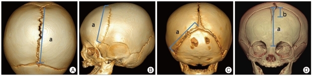
Fig. 1 Length measurement of the cranial suture. A : Total length of sagittal suture in this case is 'a' and length of closed suture line is 0. B : Total length of coronal suture in this case is 'a' and length of closed suture line is also 'a'. C : Total length of lambdoidal suture. D: Total length of metopic suture in this case is 'a' and length of closed suture line is 'a-b'.

Fig. 2 Suture closure grade in coronal suture. A : Grade 0=no closure along the whole length. B : Grade 1=partial or intermittent closure. C : Grade 2=complete closure (with a visible suture line). D : Grade 3=complete ossification (with no visible trace of the suture line).
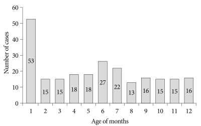
Fig. 3 Graph showing age distribution of the infants. The infants are almot evenly distributed over the different age groups after one month of age.
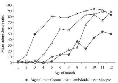
Fig. 4 Differences in mean suture closure rates of major cranial sutures by age of months. Sagittal suture is all completely patent during first 4 month in all cases, then it start to partially obliterated since 5 months. At the age of 12 months, mean suture closure rates is 49.5%. In coronal and lambdoidal suture, suture closure rates within one month of age are 1.9% and 2.6%, respectively, then progressively increased during one year, mean suture closure rates at the age of 12 months is 86.8% and 88.9%, respectively. Suture closure rates of metopic suture are 1.9% within one month of age but at the age of 3 months, about 50% of entire metopic sutures are closed. At the 11 months of age, mean suture closure rates is 91.5%.
Cited by 1 articles
-
Craniofacial malformation treatment: craniosynostosis and positional plagiocephaly
Dong Ha Park, Soo Han Yoon
J Korean Med Assoc. 2012;55(9):878-886. doi: 10.5124/jkma.2012.55.9.878.
Reference
-
1. Albin RE, Hendee RW Jr, O'Donnell RS, Majure JA. Trigonocephaly : refinements in reconstruction. Experience with 33 patients. Plast Reconstr Surg. 1985; 76:202–211. PMID: 4023093.2. Baer MJ. Patterns of growth of the skull as revealed by vital staining. Hum Biol. 1954; 26:80–126. PMID: 13191803.3. Choudhary AK, Jha B, Boal DK, Dias M. Occipital sutures and its variations : the value of 3D-CT and how to differentiate it from fractures using 3D-CT? Surg Radiol Anat. 2010; 32:807–816. PMID: 20174986.
Article4. Chugani HT. Functional brain imaging in pediatrics. Pediatr Clin North Am. 1992; 39:777–799. PMID: 1635806.
Article5. Cohen MM Jr. Etiopathogenesis of craniosynostosis. Neurosurg Clin N Am. 1991; 2:507–513. PMID: 1821298.
Article6. Cohen MM Jr. Sutural biology and the correlates of craniosynostosis. Am J Med Genet. 1993; 47:581–616. PMID: 8266985.
Article7. Fearon JA, Kolar JC, Munro IR. Trigonocephaly-associated hypotelorism : is treatment necessary? Plast Reconstr Surg. 1996; 97:503–509. discussion 510-511. PMID: 8596780.8. Furuya Y, Edwards MS, Alpers CE, Tress BM, Norman D, Ousterhout DK. Computerized tomography of cranial sutures. Part 2 : abnormalities of sutures and skull deformity in craniosynostosis. J Neurosurg. 1984; 61:59–70. PMID: 6726412.9. Harth S, Obert M, Ramsthaler F, Reuss C, Traupe H, Verhoff MA. Ossification degrees of cranial sutures determined with flat-panel computed tomography : narrowing the age estimate with extrema. J Forensic Sci. 2010; 55:690–694. PMID: 20345795.
Article10. Kirmi O, Lo SJ, Johnson D, Anslow P. Craniosynostosis : a radiological and surgical perspective. Semin Ultrasound CT MR. 2009; 30:492–512. PMID: 20099636.11. Madeline LA, Elster AD. Suture closure in the human chondrocranium : CT assessment. Radiology. 1995; 196:747–756. PMID: 7644639.
Article12. Manzanares MC, Goret-Nicaise M, Dhem A. Metopic sutural closure in the human skull. J Anat. 1988; 161:203–215. PMID: 3254893.13. Marsh JL, Jenny A, Galic M, Picker S, Vannier MW. Surgical management of sagittal synostosis. A quantitative evaluation of two techniques. Neurosurg Clin N Am. 1991; 2:629–640. PMID: 1821309.14. Mathijssen IM, Vaandrager JM, van der Meulen JC, Pieterman H, Zonneveld FW, Kreiborg S, et al. The role of bone centers in the pathogenesis of craniosynostosis: an embryologic approach using CT measurements in isolated craniosynostosis and Apert and Crouzon syndromes. Plast Reconstr Surg. 1996; 98:17–26. PMID: 8657773.
Article15. Morriss-Kay GM, Wilkie AO. Growth of the normal skull vault and its alteration in craniosynostosis : insights from human genetics and experimental studies. J Anat. 2005; 207:637–653. PMID: 16313397.
Article16. Opperman LA. Cranial sutures as intramembranous bone growth sites. Dev Dyn. 2000; 219:472–485. PMID: 11084647.
Article17. Parisi M, Mehdizadeh HM, Hunter JC, Finch IJ. Evaluation of craniosynostosis with three-dimensional CT imaging. J Comput Assist Tomogr. 1989; 13:1006–1012. PMID: 2584477.
Article18. Park SH, Yoon DY, Lee KH, Oh SM. Three-dimensional CT Imaging in the Evaluation of Craniosynostosis. J Korean Neurosurg Soc. 1996; 25:1468–1472.19. Pollack IF, Losken HW, Fasick P. Diagnosis and management of posterior plagiocephaly. Pediatrics. 1997; 99:180–185. PMID: 9024443.
Article20. Sze RW, Hopper RA, Ghioni V, Gruss JS, Ellenbogen RG, King D, et al. MDCT diagnosis of the child with posterior plagiocephaly. AJR Am J Roentgenol. 2005; 185:1342–1346. PMID: 16247160.
Article21. Van Ooijen PM, Bongaerts AH, Witkamp R, Wijker A, Tukker W, Oudkerk M. Multi-detector computed tomography and 3-dimensional imaging in a multi-vendor picture archiving and communications systems (PACS) environment. Acad Radiol. 2004; 11:649–660. PMID: 15172367.
Article22. Vannier MW, Pilgram TK, Marsh JL, Kraemer BB, Rayne SC, Gado MH, et al. Craniosynostosis : diagnostic imaging with three-dimensional CT presentation. AJNR Am J Neuroradiol. 1994; 15:1861–1869. PMID: 7863936.23. Virapongse C, Shapiro R, Sarwar M, Bhimani S, Crelin ES. Computed tomography in the study of the development of the skull base : 1. Normal morphology. J Comput Assist Tomogr. 1985; 9:85–94. PMID: 3968285.
- Full Text Links
-
- Actions
-
Cited
- CITED
-
- Close
- Share
-
- Similar articles
-
- In vitro Fusion of the Posterior Frontal Calvarial Suture in the Mouse
- The pathogenesis of craniosynostosis in the fetus
- Craniofacial growth following unilateral lambdoid synostosis in rabbits
- Isolation and Characterization of Human Suture Mesenchymal Stem Cells In Vitro
- Growth and Development in Infants and Children Born Prematurely Who were Registered at the Public Health Center in G City
