Arthroscopic Reduction and Percutaneous Fixation of Scaphoid Fracture and Nonunion
- Affiliations
-
- 1Department of Orthopaedic Surgery, Soonchunhyang University College of Medicine, Bucheon, Korea. kbsos@schbc.ac.kr
- KMID: 2186516
- DOI: http://doi.org/10.4055/jkoa.2007.42.4.530
Abstract
-
PURPOSE: To analyze the results of an arthroscopic reduction and percutaneous fixation of scaphoid fracture and nonunion.
MATERIALS AND METHODS
Fourteen scaphoid fractures or nonunion patients were analyzed clinically. There were 13 men and 1 woman, with a mean age of 30 (14-45) years. The average follow-up time was 13 months (12-18). Three cases had delayed union, 5 cases had nonunion and 6 cases had a fracture. After fluoroscopic reduction and an arthroscopic examination, the scaphoid was fixed with an Acutrak screw or K-wire percutaneously. The serial radiographs were checked by 2 weeks to confirm the bony union. The Mayo wrist score and DASH were used to assess the functional result at the final follow up.
RESULTS
Bony union was acquired in 13 cases in a mean time of 8 weeks. The mean Mayo wrist score was 86 points (60-100) with 7 excellent, 4 good, 2 fair and 1 poor case. The mean DASH was 11.1 points (0-63.3).
CONCLUSION
In scaphoid fracture and delayed union, good results could be obtained by an arthroscopic reduction and percutaneous fixation. However, in scaphoid nonunion, this technique should only be used in selected cases.
Keyword
Figure
-

Fig. 1 (A) With fluoroscopic guidance, after initially extending the wrist, a volar guide-wire was driven in a volar to dorsal direction after reducing the fracture. Next the wrist was flexed and a dorsal guide-wire was driven in the dorsal to volar direction. (B) An arthroscopic examination includes visualization of the fracture gap and integrity of the interosseous ligament. (C) A standard cannulated Acutrak screw, 4 mm shorter than the length of the scaphoid, was inserted.
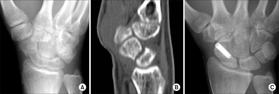
Fig. 2 (A) Oblique radiograph of an 18-year old man shows scaphoid nonunion. (B) CT shows bone resorption and a gap at the scaphoid waist. (C) Postoperative 12 months posteroanterior radiograph of the wrist shows bony union and a volarly inserted screw.

Fig. 3 (A) Oblique radiograph of 20-year old man shows scaphoid delayed union. (B) CT shows a gap at the scaphoid waist. (C) Postoperative 12 months posteroanterior radiograph of the wrist shows bony union and a dorsally inserted screw.
Reference
-
1. Adams BD, Blair WF, Reagan DS, Grundberg AB. Technical factors related to Herbert screw fixation. J Hand Surg Am. 1988. 13:893–899.
Article2. Filan SL, Herbert TJ. Herbert screw fixation of scaphoid fractures. J Bone Joint Surg Br. 1996. 78:519–529.
Article3. Garcia-Elias M, Vall A, Salo JM, Lluch AL. Carpal alignment after different surgical approaches to the scaphoid: a comparative study. J Hand Surg Am. 1988. 13:604–612.
Article4. Haddad FS, Goddard NJ. Acute percutaneous scaphoid fixation. A pilot study. J Bone Joint Surg Br. 1998. 80:95–99.5. Herbert TJ. Open volar repair of acute scaphoid fractures. Hand Clin. 2001. 17:589–599.
Article6. Hudak PL, Amadio PC, Bombardier C. Development of an upper extremity outcome measure: the DASH (disabilities of the arm, shoulder, and hand) [corrected]. The Upper Extremity Collaborative Group (UECG). Am J Ind Med. 1996. 29:602–608.7. Kamineni S, Lavy CB. Percutaneous fixation of scaphoid fractures. An anatomical study. J Hand Surg Br. 1999. 24:85–88.8. Lim JY, Lee HY, Song JH, Kang JW, Lee JY. Evaluation of the reliability, construct validity, and responsiveness of the Korean version of the DASH. J Korean Soc Surg Hand. 2005. 10:192–198.9. Rettig AC, Kollias SC. Internal fixation of acute stable scaphoid fractures in the athlete. Am J Sports Med. 1996. 24:182–186.
Article10. Rettig ME, Raskin KB. Retrograde compression screw fixation of acute proximal pole scaphoid fractures. J Hand Surg Am. 1999. 24:1206–1210.
Article11. Shih JT, Lee HM, Hou YT, Tan CM. Results of arthroscopic reduction and percutaneous fixation for acute displaced scaphoid fractures. Arthroscopy. 2005. 21:620–626.
Article12. Slade JF, Dodds SD. Minimally invasive management of scaphoid nonunions. Clin Orthop Relat Res. 2006. 445:108–119.
Article13. Slade JF 3rd, Geissler WB, Gutow AP, Merrell GA. Percutaneous internal fixation of selected scaphoid nonunions with an arthroscopically assisted dorsal approach. J Bone Joint Surg Am. 2003. 85:Suppl 4. 20–32.
Article14. Slade JF 3rd, Grauer JN, Mahoney JD. Arthroscopic reduction and percutaneous fixation of scaphoid fractures with a novel dorsal technique. Orthop Clin North Am. 2001. 32:247–261.
Article15. Slade JF, Taksali S, Safanda J. Combined fractures of the scaphoid and distal radius: a revised treatment rationale using percutaneous and arthroscopic techniques. Hand Clin. 2005. 21:427–441.
Article16. Sung IH, Jung JH, Lee KH, Choi CH, Yoon JP. Palmar percutaneous cannulated Herbert-Whipple screw fixation of scaphoid fractures. J Korean Soc Surg Hand. 2005. 10:129–135.17. Taras JS, Sweet S, Shum W, Weiss LE, Bartolozzi A. Percutaneous and arthroscopic screw fixation of scaphoid fractures in the athlete. Hand Clin. 1999. 15:467–473.
Article18. Trumble TE, Gilbert M, Murray LW, Smith J, Rafijah G, McCallister WV. Displaced scaphoid fractures treated with open reduction and internal fixation with a cannulated screw. J Bone Joint Surg Am. 2000. 82:633–641.
Article19. Trumble TE, Clarke T, Kreder HJ. Non-union of the scaphoid. Treatment with cannulated screws compared with treatment with Herbert screws. J Bone Joint Surg Am. 1996. 78:1829–1837.
Article20. Whipple TL. The role of arthroscopy in the treatment of intra-articular wrist fractures. Hand Clin. 1995. 11:13–18.
Article21. Yip HS, Wu WC, Chang RY, So TY. Percutaneous cannulated screw fixation of acute scaphoid waist fracture. J Hand Surg Br. 2002. 27:42–46.
Article
- Full Text Links
-
- Actions
-
Cited
- CITED
-
- Close
- Share
-
- Similar articles
-
- Arthroscopic Bone Grafting and Kirschner-Wires Fixation for Scaphoid Nonunion
- Arthroscopic Bone Grafting and Percutaneous K-Wires Fixation for the Treatment of Scaphoid Nonunion: Surgical Technique
- Scaphoid Fractures and Nonunions: Recent Trends of Treatment
- Scaphoid Fractures and Nonunion
- Headless Autocompression Screw Fixation of Scaphoid Fractures Using Open Dorsal Approach
