Subdeltoid Bursa Chondromatosis Associated with Osteochondral Lesion of the Proximal Humerus and a Rotator Cuff Tear
- Affiliations
-
- 1Department of Orthopedic Surgery, Haeundae Paik Hospital, Busan, Korea.
- 2Department of Orthopedic Surgery, Ilsan Paik Hospital, Inje University College of Medicine, Koyang, Korea.
- 3Department of Orthopedic Surgery, Seoul Nanoori Hospital, Seoul, Korea. tsnam74@gmail.com
- KMID: 2185465
- DOI: http://doi.org/10.4055/jkoa.2011.46.3.256
Abstract
- Synovial chondromatosis involving the bursa is uncommon, and those cases synovial chondromatosis within the bursa around the shoulder are especially rare. We report here a case of a 57-year-old male who had subdeltoid bursal subdeltoid bursal chondromatosis associated with osteochondral lesion of the proximal humerus and a rotator cuff tear. We also review the relevant literatures.
Figure
-
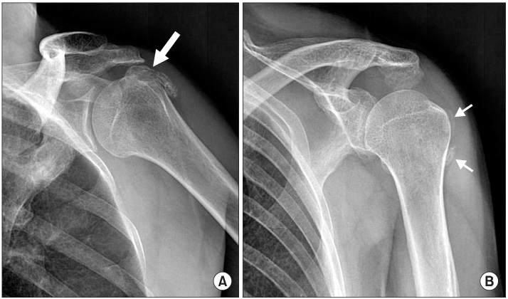
Figure 1 The left shoulder AP (A) and 30 degree caudal tilt (B) radiographs show oval shaped calcific loose bodies in the subdeltoid recess (thick arrow) and bony protrusion on greater tuberosity of the humerus (thin arrows).
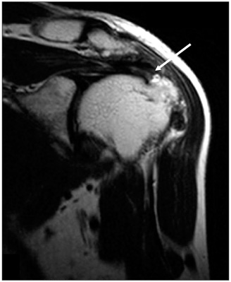
Figure 2 T2-weighted coronal MR image of the left shoulder shows nearly full-thickness tear of supraspinatus tendon (arrow).
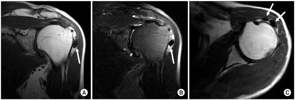
Figure 3 T1-weighted coronal (A), T2-weighted coronal (B) and axial (C) MR images of the left shoulder show two loose bodies in the subdeltoid recess (arrows) and bony protrusion on greater tuberosity of the humerus (asterix).
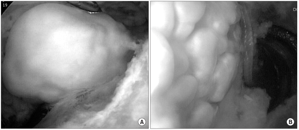
Figure 4 Arthroscopic findings show a large chondral loose body in subdeltoid recess (A) and cobble-stone appearance cartilage formation on the lateral side of the humeral head (B).
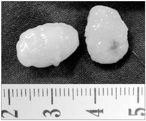
Figure 5 Gross specimen of loose bodies.

Figure 6 Arthroscopic findings show tear of supraspinatus (A) and repaired supraspinatus after surgery (B).

Figure 7 Microscopic finding of synovial loose body demonstrates cartilaginous metaplasia (thick arrow) beneath synovial surface (thin arrow) (Hematoxylin-Eosin stain, original magnification ×150).
Reference
-
1. Volpin G, Nerubay J, Oliver S, Katznelson A. Synovial osteochondromatosis of the shoulder joint. Am Surg. 1980. 46:422–424.2. Ogawa K, Takahashi M, Inokuchi W. Bilateral osteochondromatosis of the subacromial bursae with incomplete rotator cuff tears. J Shoulder Elbow Surg. 1999. 8:78–81.
Article3. Milgram JW, Hadesman WM. Synovial osteochondromatosis in the subacromial bursa. Clin Orthop Relat Res. 1988. (236):154–159.
Article4. Symeonides P. Bursal chondromatosis. J Bone Joint Surg Br. 1966. 48:371–373.
Article5. Huang TF, Wu JJ, Chen TS. Bilateral shoulder bursal osteochondromatosis associated with complete rotator cuff tear. J Shoulder Elbow Surg. 2004. 13:108–111.
Article6. Bruggeman NB, Sperling JW, Shives TC. Arthroscopic technique for treatment of synovial chondromatosis of the glenohumeral joint. Arthroscopy. 2005. 21:633.
Article7. Iwata H, Ono S, Sato K, Sato T, Kawamura M. Bone morphogenetic protein-induced muscle- and synovium-derived cartilage differentiation in vitro. Clin Orthop Relat Res. 1993. (296):295–300.
Article8. Small R, Jaffe WL. Tenosynovial chondromatosis of the shoulder. Bull Hosp Jt Dis Orthop Inst. 1981. 41:37–47.9. Ko JY, Wang JW, Chen WJ, Yamamoto R. Synovial chondromatosis of the subacromial bursa with rotator cuff tearing. J Shoulder Elbow Surg. 1995. 4:312–316.10. Sim FH, Dahlin DC, Ivins JC. Extra-articular synovial chondromatosis. J Bone Joint Surg Am. 1977. 59:492–495.
Article
- Full Text Links
-
- Actions
-
Cited
- CITED
-
- Close
- Share
-
- Similar articles
-
- Reverse Total Shoulder Replacement for an Enchondroma with Concomitant Rotator Cuff Tear Arthropathy: A Case Report
- Comparison of Ultrasonographic and Arthrographic Findings according to the Severity of the Rotator Cuff Tear
- Reverse Shoulder Arthroplasty for Humeral Head Fracture with Massive Rotator Cuff Tear in Elderly Patient
- Delaminated Rotator Cuff Tear: Concurrent Concept and Treatment
- A Retrospective Analysis of the Relationship Between Rotator Cuff Tear and Biceps Lesion
