Sarcoidosis Presenting as Knee Pain
- Affiliations
-
- 1Department of Orthopedic Surgery, Bucheon Hospital, Bucheon, Korea. hwangseokha@naver.com
- 2Department of Orthopedic Surgery, Seoul Hospital, Soon Chun Hyang University College of Medicine, Seoul, Korea.
- KMID: 2185378
- DOI: http://doi.org/10.4055/jkoa.2012.47.4.299
Abstract
- Sarcoidosis is a multisystem granulomatous disease of unknown etiology with variable manifestations, which may affect virtually any organ. Muscular sarcoidosis is a rare entity, and among this group of muscular lesions, the mass-like muscular sarcoidosis type is extremely rare. We reported a muscular sarcoidosis case that presented with right knee pain and described the clinical results with an added review of the relevant literature.
Keyword
MeSH Terms
Figure
-
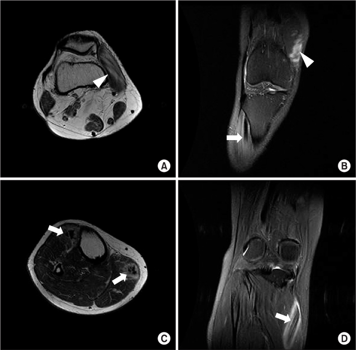
Figure 1 (A, B) An axial and coronal T2-weighted magnetic resonance (MR) images show hyperintensity (white arrow head) in the vastus medialis muscle fiber. (C) An axial T2-weighted MR image shows ovoid nodules (white arrows) scattered throughout muscles of the lower extremities (dark star sign). (D) T2-weighted coronal MR image shows an inner stripe of decreased signal intensity and outer stripes of significantly increased signal intensity in the tibialis anterior muscle (B, white arrow) and the proximal medial gastrocnemius muscle (three stripe sign, white arrow).
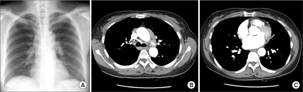
Figure 2 (A) A chest radiograph shows perihilar nodular densities suggesting bihilar lymphadenopathy. (B, C) Dynamic chest computed tomography shows homogeneously enhancing enlarged lymph nodes at both lower paratrachea, both hila and subcarina.
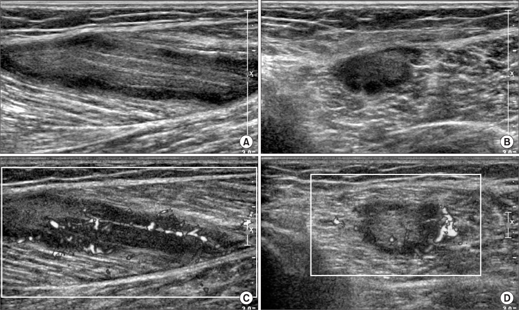
Figure 3 (A, B) Longitudinal and short axial scans through the right medial gastrocnemius muscle show an inner structure of increased echogenicity and outer area of decreased echogenicity. (C, D) Longitudinal and short axial color doppler sonographies show plenty of vascularity in the proximal medial gastrocnemius muscle.
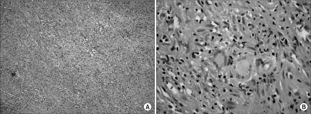
Figure 4 (A) Numerous epitheloid cells formed noncaseating granulomas (H&E, ×40). (B) A histopathologic specimen of the needle aspiration showed numerous epithelioid histiocytes arranged in sheets and a few multinucleated giants cells of Langhans-type (H&E, ×200).
Reference
-
1. Zisman DA, Biermann JS, Martinez FJ, Devaney KO, Lynch JP 3rd. Sarcoidosis presenting as a tumorlike muscular lesion. Case report and review of the literature. Medicine (Baltimore). 1999. 78:112–122.
Article2. Fayad F, Lioté F, Berenbaum F, Orcel P, Bardin T. Muscle involvement in sarcoidosis: a retrospective and followup studies. J Rheumatol. 2006. 33:98–103.3. Baughman RP, Lower EE, du Bois RM. Sarcoidosis. Lancet. 2003. 361:1111–1118.
Article4. Ogane K, Kato T, Mizushima I, Kawano M, Yamagishi M. A case of sarcoidosis developing as sarcoid myopathy concomitant with systemic sclerosis and review of the literature. Mod Rheumatol. 2012. 22:142–146.
Article5. Kolilekas L, Triantafillidou C, Manali E, Rontogianni D, Chatziioannou S, Papiris S. The many faces of sarcoidosis: asymptomatic muscle mass mimicking giant-cell tumor. Rheumatol Int. 2009. 29:1389–1390.
Article6. Otake S. Sarcoidosis involving skeletal muscle: imaging findings and relative value of imaging procedures. AJR Am J Roentgenol. 1994. 162:369–375.
Article7. Yamamoto T, Nagira K, Akisue T, et al. Aspiration biopsy of nodular sarcoidosis of the muscle. Diagn Cytopathol. 2002. 26:109–112.
Article8. Chen HH, Hsieh TY, Chen DY, Lan HHC, Hsieh CW. Sonographic features of nodular-type muscular sarcoidosis. J Med Ultrasound. 2007. 15:197–201.
Article9. Torralba KD, Quismorio FP Jr. Sarcoidosis and the rheumatologist. Curr Opin Rheumatol. 2009. 21:62–70.
Article10. Guis S, Mattéi JP, Lioté F. Drug-induced and toxic myopathies. Best Pract Res Clin Rheumatol. 2003. 17:877–907.
Article
- Full Text Links
-
- Actions
-
Cited
- CITED
-
- Close
- Share
-
- Similar articles
-
- Systemic Sarcoidosis Presenting with Arrhythmia
- A Case of Muscular Sarcoidosis diagnosed by Gallium-67 Scintigraphy and Magnetic Resonance Imaging
- Scar Sarcoidosis after Blepharoplasty: A Case Series
- Sarcoidosis Initially Presenting as a Nasal Cavity Mass Misdiagnosed as Tuberculosis
- Psoriasiform Sarcoidosis
