Metastatic Osteosarcoma to the Breast Presenting as a Densely Calcified Mass on Mammography
- Affiliations
-
- 1Department of Radiology, Severance Hospital, Research Institute of Radiological Science, Yonsei University College of Medicine, Seoul, Korea. lvjenny@yuhs.ac
- 2Department of Pathology, Yonsei University College of Medicine, Seoul, Korea.
- KMID: 2176326
- DOI: http://doi.org/10.4048/jbc.2016.19.1.87
Abstract
- Osteosarcoma most commonly metastasizes to the lung or the skeleton, and metastatic osteosarcoma to the breast is very rare, with only a few cases reported. Due to its rarity, little has been reported about its imaging features. In this report, we represent a 58-year-old woman with metastatic osteosarcoma to the right breast from a tibial osteosarcoma. The imaging features of the metastatic osteosarcoma to the breast by using dedicated breast imaging modalities are described. Although rare, metastatic osteosarcoma to the breast should be considered when dense calcified masses with suspicious features are seen on breast imaging in patients with a history of osteosarcoma.
Keyword
MeSH Terms
Figure
-
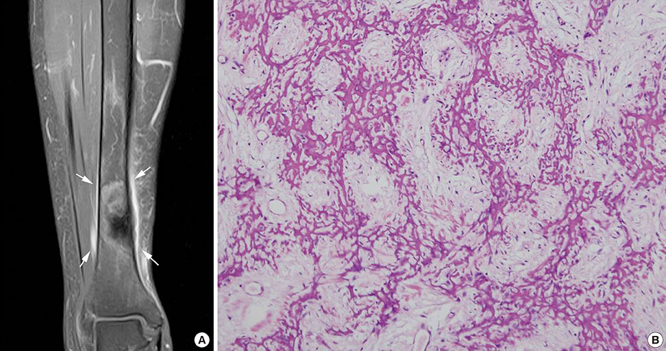
Figure 1 Magnetic resonance imaging and pathologic features of the osteosarcoma involving the right tibia. (A) A 4.8-cm-sized osteosclerotic mass (arrows) with periosteal reaction is seen in the diaphysis of the right distal tibia. (B) This mass was diagnosed as conventional osteosarcoma, osteoblastic type, showing lacy architectural pattern on excisional biopsy (H&E stain, ×200).
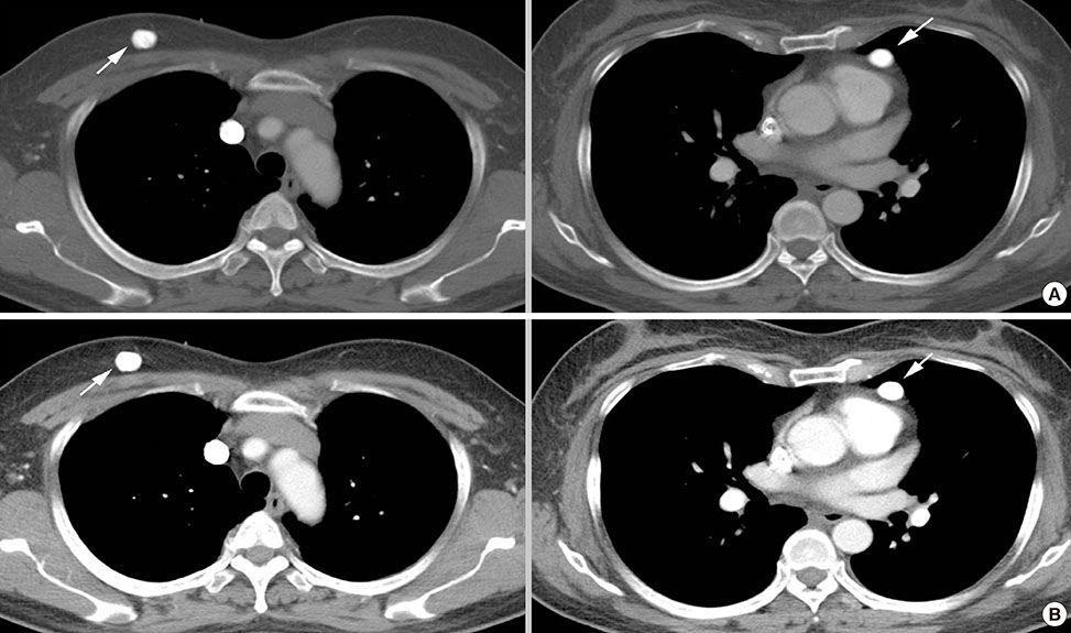
Figure 2 Metastatic osteosarcoma detected on chest computed tomography (CT). (A) Two calcified masses were seen on initial chest CT, one in the right breast, and one at the anterior mediastinum (arrows). (B) Follow-up chest CT performed 3 weeks later revealed size increase of these masses, breast mass from 11 to 14 mm and mediastinal mass from 12 to 15 mm (arrows), raising the suspicion for metastasis.
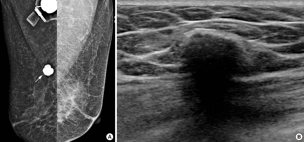
Figure 3 Mammography and breast ultrasonography (US) examinations of the right breast mass. (A) Mediolateral oblique views of mammography show a dense calcified mass (arrow) with spiculated margins in the right upper breast. (B) Breast US performed on the same day shows a 1.5-cm dense calcified mass with posterior shadowing. Percutaneous biopsy was considered difficult due to the dense calcifications, and the patient underwent excisional biopsy.
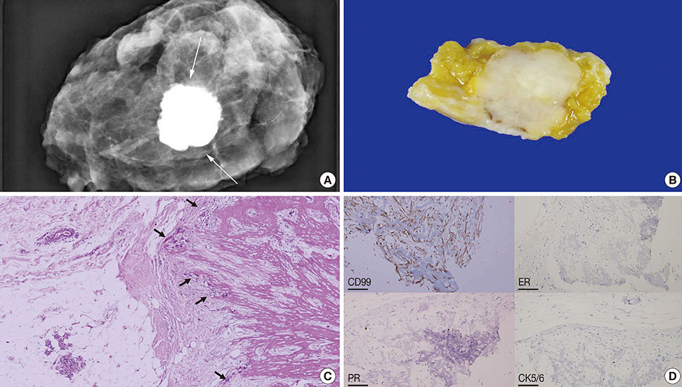
Figure 4 Specimen mammography and the pathology features of the right breast mass. (A) Specimen mammography reveals the dense calcified mass (arrows) seen on mammography in the right breast, confirming complete excision of the mass. (B) Similar features are seen on gross specimen, which reveals a dense calcified mass within the breast tissue. (C) Microscopic examination shows a mass with predominant osteoid matrix production (arrows) (H&E stain, ×40). (D) Immunohistochemically, these tumor cells were positive for cluster of differentiation 99 (CD99), negative for estrogen receptor (ER), progesterone receptor (PR), and cytokeratin 5/6 (CK5/6). These findings are consistent with those of metastatic osteosarcoma to the breast, rather than primary metaplastic carcinoma of the breast (×100).
Cited by 1 articles
-
Invasive Breast Cancer Presenting as a Mass Replaced by Calcification on Mammography: A Report of Two Cases
Joo Hee Jeun, Jin Hwa Lee, Eun Cho, Su Jin Kim, Eun Hwa Park, Kyung Do Byun
J Korean Soc Radiol. 2019;80(3):591-597. doi: 10.3348/jksr.2019.80.3.591.
Reference
-
1. Yeh CN, Lin CH, Chen MF. Clinical and ultrasonographic characteristics of breast metastases from extramammary malignancies. Am Surg. 2004; 70:287–290.2. Mun SH, Ko EY, Han BK, Shin JH, Kim SJ, Cho EY. Breast metastases from extramammary malignancies: typical and atypical ultrasound features. Korean J Radiol. 2014; 15:20–28.
Article3. Basu S, Moghe SH, Shet T. Metastasis of humeral osteosarcoma to the contralateral breast detected by 99mTc-MDP skeletal scintigraphy. Jpn J Radiol. 2009; 27:455–457.
Article4. Vieira SC, Tavares MB, da Cruz Filho AJ, Sousa RB, Dos Santos LG. Metastatic osteosarcoma to the breast: a rare case. J Obstet Gynaecol Res. 2010; 36:891–893.
Article5. Chan RS, Kumar G, Vijayananthan AA. Rare occurrence of bilateral breast and peritoneal metastases from osteogenic sarcoma. Singapore Med J. 2013; 54:e68–e71.
Article6. Roebuck DJ, Sato JK, Fahmy J. Breast metastasis in osteosarcoma. Australas Radiol. 1999; 43:108–110.
Article7. Kim SJ, Choi JA, Lee SH, Choi JY, Hong SH, Chung HW, et al. Imaging findings of extrapulmonary metastases of osteosarcoma. Clin Imaging. 2004; 28:291–300.
Article8. Salah S, Ahmad R, Sultan I, Yaser S, Shehadeh A. Osteosarcoma with metastasis at initial diagnosis: current outcomes and prognostic factors in the context of a comprehensive cancer center. Mol Clin Oncol. 2014; 2:811–816.
Article9. Surov A, Fiedler E, Holzhausen HJ, Ruschke K, Schmoll HJ, Spielmann RP. Metastases to the breast from non-mammary malignancies: primary tumors, prevalence, clinical signs, and radiological features. Acad Radiol. 2011; 18:565–574.10. Momoi H, Wada Y, Sarumaru S, Tamaki N, Gomi T, Kanaya S, et al. Primary osteosarcoma of the breast. Breast Cancer. 2004; 11:396–400.
Article11. Dragoumis D, Bimpa K, Assimaki A, Tsiftsoglou A. Primary osteogenic sarcoma of the breast. Singapore Med J. 2008; 49:e315–e317.12. Trihia H, Valavanis C. Histopathology and molecular pathology of bone and extraskeletal osteosarcomas. In : Agarwal M, editor. Osteosarcoma. Rijeka: InTech;2012.13. Leibl S, Gogg-Kammerer M, Sommersacher A, Denk H, Moinfar F. Metaplastic breast carcinomas: are they of myoepithelial differentiation? Immunohistochemical profile of the sarcomatoid subtype using novel myoepithelial markers. Am J Surg Pathol. 2005; 29:347–353.
Article14. Geller DS, Gorlick R. Osteosarcoma: a review of diagnosis, management, and treatment strategies. Clin Adv Hematol Oncol. 2010; 8:705–718.15. Bielack S, Carrle D, Casali PG. ESMO Guidelines Working Group. Osteosarcoma: ESMO clinical recommendations for diagnosis, treatment and follow-up. Ann Oncol. 2009; 20:Suppl 4. 137–139.
Article
- Full Text Links
-
- Actions
-
Cited
- CITED
-
- Close
- Share
-
- Similar articles
-
- Mucinous Breast Carcinoma Presenting as a Coarse and Densely Calcified Mass on Mammography: A Case Report
- Primary osteosarcoma of the breast: a case report
- Primary Osteosarcoma of the Breast: A case report
- Bladder Cancer Metastasis to the Breast in a Male Patient: Imaging Findings on Mammography and Ultrasonography
- Misdiagnosed Breast Cancer on Mammography Retrospective Analysis in 17 Cases
