J Gynecol Oncol.
2011 Dec;22(4):295-298. 10.3802/jgo.2011.22.4.295.
Paratubal serous borderline tumor
- Affiliations
-
- 1Department of Obstetrics and Gynecology, Asan Medical Center, University of Ulsan College of Medicine, Seoul, Korea. catgut1-0@hanmail.net
- 2Department of Pathology, Asan Medical Center, University of Ulsan College of Medicine, Seoul, Korea.
- KMID: 2173633
- DOI: http://doi.org/10.3802/jgo.2011.22.4.295
Abstract
- Although paratubal cysts are well-characterized incidental findings, paratubal serous borderline tumors are very rare, with only one case report in the literature. We describe here a 27-year-old, nulliparous, married woman with a paratubal serous borderline tumor. The patient presented with a huge pelvic mass accompanied by flank pain and underwent paratubal cystectomy and fertility-sparing surgical staging procedures. Thirteen months after surgery, she delivered a healthy baby at term. She is well, without evidence of disease, 20 months after surgery. Because paratubal serous borderline tumors are very rare, their optimal management must be extrapolated from their ovarian counterparts.
Figure
-

Fig. 1 Transvaginal ultrasonography (A) and abdomino-pelvic computed tomography (B) showing a 16 cm cystic mass with enhancing intramural solid nodules (arrows) in the pelvic cavity.
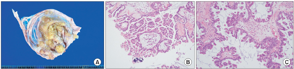
Fig. 2 (A) Grossly, the tumor had a homogeneous purplish white inner surface and multiple yellow papillary projections up to 2.1 cm in greatest dimension. (B, C) Microscopic finding of paratubal serous borderline tumor: epithelial cells with mild nuclear atypia show stratification and appear to be free-floating around the smaller papillae (B, C: H&E, ×400).
Reference
-
1. Villella JA, Pauli SA, Wang J, Intengan M, Lele S. Tumors of low malignant potential arising in the fallopian tube: case reports. Eur J Gynaecol Oncol. 2005. 26:327–329.2. Seamon LG, Holt CN, Suarez A, Richardson DL, Carlson MJ, O'Malley DM. Paratubal borderline serous tumors. Gynecol Oncol. 2009. 113:83–85.3. Scully RE, Young RH, Clement PB. Tumors of the ovary, maldeveloped gonads, fallopian tube and broad ligament. 1998. Washington, DC: American Registry of Pathology, Armed Forces Institute of Pathology.4. Gatto V, Selim MA, Lankerani M. Primary carcinoma of the fallopian tube in an adolescent. J Surg Oncol. 1986. 33:212–214.5. Valerdiz Casasola S, Pardo Mindan J. Cystadenofibroma of fallopian tube. Appl Pathol. 1989. 7:256–259.6. Zheng W, Wolf S, Kramer EE, Cox KA, Hoda SA. Borderline papillary serous tumour of the fallopian tube. Am J Surg Pathol. 1996. 20:30–35.7. Alvarado-Cabrero I, Navani SS, Young RH, Scully RE. Tumors of the fimbriated end of the fallopian tube: a clinicopathologic analysis of 20 cases, including nine carcinomas. Int J Gynecol Pathol. 1997. 16:189–196.8. Kayaalp E, Heller DS, Majmudar B. Serous tumor of low malignant potential of the fallopian tube. Int J Gynecol Pathol. 2000. 19:398–400.9. Haratz-Rubinstein N, Fromberg E, Lederman S. Sonographic diagnosis of a serous tumor of low malignant potential of the fallopian tube. J Ultrasound Med. 2004. 23:869–872.10. Krasevic M, Stankovic T, Petrovic O, Smiljan-Severinski N. Serous borderline tumor of the fallopian tube presented as hematosalpinx: a case report. BMC Cancer. 2005. 5:129.11. Park JH, Lim MC, Kim SW, Kim SK, Yoo CW, Park SY. A serous borderline tumor of the fallopian tube detected incidentally. Korean J Obstet Gynecol. 2009. 52:1045–1050.12. Abreu R, Dick M, Simoes Silva T, Mota F, Bettencourt E. Serous borderline tumor of the fallopian tube presented as an adnexal mass. Arch Gynecol Obstet. 2011. 283:349–352.
- Full Text Links
-
- Actions
-
Cited
- CITED
-
- Close
- Share
-
- Similar articles
-
- Serous borderline tumor of the fallopian tube
- Two Cases of Advanced Ovarian Serous Tumor of Borderline Malignancy
- A Serous Papillary Cystadenoma of Borderline Malignancy in Testis
- Ovarian Borderline Epithelial Tumors
- A Case of Stage III c Borderline Malignant Ovarian Surface Papilloma with Invasive Peritoneal Implant
