Perils of Diagnosis and Detection of Subungual Squamous Cell Carcinoma
- Affiliations
-
- 1Department of Surgery, Division of Plastic Surgery, UMDNJ-New Jersey Medical School, New Jersey, USA. fleeglea@umdnj.edu
- 2Department of Pathology and Laboratory Medicine, UMDNJ-New Jersey Medical School, New Jersey, USA.
- KMID: 2171852
- DOI: http://doi.org/10.5021/ad.2011.23.S3.S285
Abstract
- Subungual squamous cell carcinoma often presents with atypical clinical manifestations, which can lead to delays in diagnosis. The presence of a tumor can be masked by the presence of infections or other misleading pathological conditions. The authors report on techniques for adequate biopsy and excision of such tumors. A case of subungual squamous cell carcinoma with invasion into the underlying bone is presented. Clinical histopathological evidence is reviewed along with human papillomavirus typing. Accurate diagnosis requires a high index of suspicion and appropriate tissue sampling.
Keyword
MeSH Terms
Figure
-
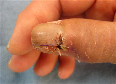
Fig. 1 Preoperative image showing scant granulomatous tissue along the radial border of the nail fold with nail deformity.

Fig. 2 High power view of a biopsy of the thumbnail fold reveals a tumor composed of whorls of an invasive proliferation of atypical keratinocytes with dyskeratosis and keratin pearls within the dermis. The tumor exhibits distinct intracellular bridges and hyperchromatic, pleomorphic nuclei. Focal areas of single cell keratinization are identified (H&E, original magnification ×400).

Fig. 3 Medium power view of the subsequent distal thumb resection shows deep involvement of the tumor reaching the cortical bone (H&E, original magnification ×100).
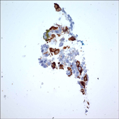
Fig. 4 Immunoperoxidase staining for cytokeratin (AE1/AE3) confirms tumor involvement within the bone. Immunohistochemistry AE1/AE3 (original magnification ×400).
Reference
-
1. Porembski MA, Rayan GM. Subungual carcinomas in multiple digits. J Hand Surg Eur Vol. 2007. 32:547–549.
Article2. High WA, Tyring SK, Taylor RS. Rapidly enlarging growth of the proximal nail fold. Dermatol Surg. 2003. 29:984–986.
Article3. Attiyeh FF, Shah J, Booher RJ, Knapper WH. Subungual squamous cell carcinoma. JAMA. 1979. 241:262–263.
Article4. Guitart J, Bergfeld WF, Tuthill RJ, Tubbs RR, Zienowicz R, Fleegler EJ. Squamous cell carcinoma of the nail bed: a clinicopathological study of 12 cases. Br J Dermatol. 1990. 123:215–222.
Article5. Stetsenko GY, McFarlane RJ, Chien AJ, Fleckman P, Swanson P, George E, et al. Subungual Bowen disease in a patient with epidermodysplasia verruciformis presenting clinically as longitudinal melanonychia. Am J Dermatopathol. 2008. 30:582–585.
Article6. Moy RL, Eliezri YD, Nuovo GJ, Zitelli JA, Bennett RG, Silverstein S. Human papillomavirus type 16 DNA in periungual squamous cell carcinomas. JAMA. 1989. 261:2669–2673.
Article7. Bogumill GP, Fleegler EJ. Tumors of the hand and upper limb. 1993. New York: Churchill Livingstone;147–149.8. Dalle S, Depape L, Phan A, Balme B, Ronger-Savle S, Thomas L. Squamous cell carcinoma of the nail apparatus: clinicopathological study of 35 cases. Br J Dermatol. 2007. 156:871–874.
Article9. Salasche SJ, Garland LD. Tumors of the nail. Dermatol Clin. 1985. 3:501–519.
Article10. Shapiro L, Baraf CS. Subungual epidermoid carcinoma and keratoacanthoma. Cancer. 1970. 25:141–152.
Article
