Ann Dermatol.
2015 Aug;27(4):394-397. 10.5021/ad.2015.27.4.394.
Button Osteoma: A Review of Ten Cases
- Affiliations
-
- 1Department of Dermatology, Kyungpook National University School of Medicine, Daegu, Korea. weonju@knu.ac.kr
- KMID: 2171495
- DOI: http://doi.org/10.5021/ad.2015.27.4.394
Abstract
- BACKGROUND
Button osteoma presents as small circumscribed ivory-like lumps on the skull vault. Although not rare, its diagnosis can be challenging for dermatologists.
OBJECTIVE
To clarify the clinical characteristics of button osteoma by reviewing 10 cases.
METHODS
Ten patients diagnosed with button osteoma at the Department of Dermatology, Kyungpook National University Hospital, between January 2011 and August 2014 were enrolled. We retrospectively reviewed medical records and analyzed demographic and clinical characteristics including sex, age, sites, number of lesions, symptoms, duration, histopathological finding, radiological findings, and treatment.
RESULTS
All patients presented with an asymptomatic small circumscribed hard lump fixed to a bony structure. There were 9 female and 1 male patient, and the mean age was 54 years (range, 28approximately61 years). The most common site was the forehead, and disease duration ranged from 2 weeks to more than 20 years. The differential diagnosis included cranial exostosis, ballooned osteoma, epidermal cyst, and lipoma. Simple radiography, ultrasonography, and computed tomography (CT) were used to make a confirmative diagnosis. Histopathological findings showed lamellated bony structures with poor vascularization. Ostectomy was performed for 5 patients, and no recurrence was detected within an average of 13.4 months after treatment.
CONCLUSION
This review characterized button osteoma. Surgical excision is a useful therapeutic modality after CT-based diagnosis. Further studies with more patients are required to confirm the findings.
Keyword
MeSH Terms
Figure
-
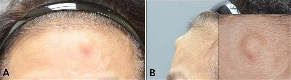
Fig. 1 A solitary well-defined asymptomatic nodule 1.3 cm in diameter is seen on the forehead. Inset: close-up view of the lesion.
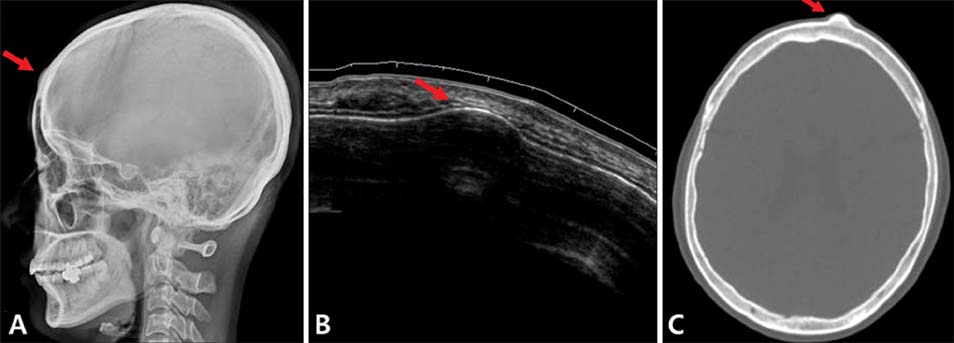
Fig. 2 (A) Simple radiography shows a well-defined smooth homogenous bony density (arrow) on the frontal bone. (B) Ultrasonography shows a 1.4 cm smooth and round bony protuberance (arrow) on the frontal bone. (C) Computed tomography shows a 1.3 cm well-defined homogenous radiopaque lesion (arrow) on the frontal area.
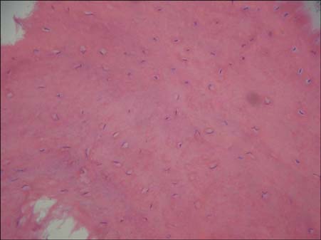
Fig. 3 Histopathological examination shows compact and mature bone with lacunae (H&E, ×200).
Reference
-
1. Haddad FS, Haddad GF, Zaatari G. Cranial osteomas: their classification and management. Report on a giant osteoma and review of the literature. Surg Neurol. 1997; 48:143–147.
Article2. Patel TR, Borah GL. Frontal bone periosteal osteomas. Plast Reconstr Surg. 2004; 114:648–651.
Article3. Eshed V, Latimer B, Greenwald CM, Jellema LM, Rothschild BM, Wish-Baratz S, et al. Button osteoma: its etiology and pathophysiology. Am J Phys Anthropol. 2002; 118:217–230.
Article4. Chun YS, Yoon KH, Choi HJ, Kim LS. Intramuscular lipoma of the frontalis muscle. Ann Dermatol. 1999; 11:98–100.
Article5. Tucker WS, Nasser-Sharif FJ. Benign skull lesions. Can J Surg. 1997; 40:449–455.6. Bracaglia R, Fortunato R, Marando A, Farallo E. Frontal exostose resection during an endoscopic subperiosteal lifting: case report. Aesthetic Plast Surg. 1997; 21:122–124.
Article7. Summers LE, Mascott CR, Tompkins JR, Richardson DE. Frontal sinus osteoma associated with cerebral abscess formation: a case report. Surg Neurol. 2001; 55:235–239.
Article8. Saraç K, Biliciler B, Vatansever M, AladaXMLLink_XYZ MA, Colak A. Unusual frontal osteoma, mimicking a haemangioma. Neuroradiology. 1996; 38:458–459.
Article9. Boffano P, Roccia F, Campisi P, Gallesio C. Review of 43 osteomas of the craniomaxillofacial region. J Oral Maxillofac Surg. 2012; 70:1093–1095.
Article10. Yamamoto T, Alvarado Gutierrez JL, Koshima I. Incisionless osteotomy for contouring the skull: pinhole osteo-chipping with irrigation for the esthetic treatment of a benign frontal osteoma. J Plast Reconstr Aesthet Surg. 2014; 67:e270–e272.
Article11. Onishi K, Maruyama Y, Sawaizumi M. Endoscopic excision of forehead osteoma. J Craniofac Surg. 1995; 6:516–518.12. Sewell LD, Adams DC, Marks VJ. Subcutaneous forehead nodules: attention to the button osteoma and frontalisassociated lipoma. Dermatol Surg. 2008; 34:791–798.
Article
