Trichoscopic Findings of Hair Loss in Koreans
- Affiliations
-
- 1Department of Dermatology, Chonbuk National University Medical School, Research Institute of Clinical Medicine of Chonbuk National University-Biomedical Research Institute of Chonbuk National University Hospital, Jeonju, Korea.
- 2Department of Dermatology, Chonnam National University Medical School, Gwangju, Korea. seongjinkim@jnu.ac.kr
- KMID: 2171454
- DOI: http://doi.org/10.5021/ad.2015.27.5.539
Abstract
- BACKGROUND
Trichoscopic findings of hair loss have been well described for the differential diagnosis of alopecia; however, critical findings were not thoroughly investigated or compared among all ethnic groups, including Asians.
OBJECTIVE
We aimed to find any characteristic trichoscopic findings in Korean alopecia patients and to verify whether those findings are closely related to previously reported observations.
METHODS
Three hundred and twenty-seven patients with hair loss of various causes and 160 normal scalps were analyzed. Trichoscopic examination was performed with a polarized-light handheld dermoscope.
RESULTS
A total of 35 patterns of trichoscopic features were represented, and certain features were significantly common or observed exclusively in a particular type of alopecia as follows: yellow dots, exclamation mark hairs, and proximal tapering hairs (alopecia areata), trichoptilosis and pointed hairs (trichotillomania), corkscrew hairs, septate hyphae hairs, and comma hairs (tinea capitis), diffuse white area, fibrotic white dots, and tufting hairs (primary cicatricial alopecia), hair diameter diversity and peripilar sign (androgenetic alopecia), and short nonvellus hairs (telogen effluvium).
CONCLUSION
The characteristic trichoscopic features for the differential diagnosis of alopecia in Koreans, shown as follicular, perifollicular, and hair shaft patterns, are similar to those of Caucasians; however, the frequencies of the pigment patterns are different between Koreans and Caucasians because of the contrast effect of the skin and hair color. Therefore, racial difference should be considered in the trichoscopic evaluation for differential diagnosis.
Keyword
MeSH Terms
Figure
-
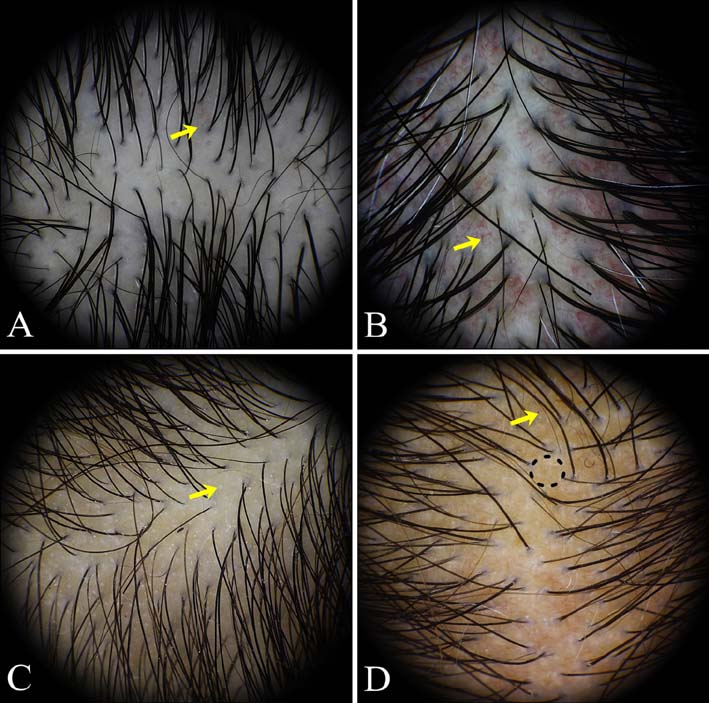
Fig. 1 Trichoscopic findings of normal subjects. (A) simple red loops (yellow arrow), (B) arborizing vessels (yellow arrow), (C) pinpoint white dots (yellow arrow), (D) dirty dots (yellow arrow), and honeycomb pigmentation (dotted circle).
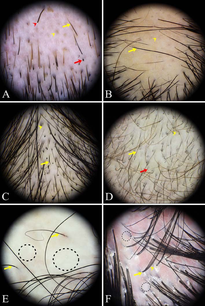
Fig. 2 Trichoscopic findings of localized hair loss. (A) Patchy alopecia areata: exclamation mark hairs (yellow arrow), yellow dots (yellow arrowhead), black dots (red arrow), broken hairs (red arrowhead). (B) Patchy alopecia areata: proximal tapering hairs (yellow arrow), short vellus hairs (yellow arrowhead). (C) Trichotillomania: trichoptilosis (yellow arrow), pointed hairs (yellow arrowhead), black dots, and broken hairs with different lengths. (D) Tinea capitis: septate hyphae hairs (yellow arrow), corkscrew hairs (yellow arrowhead), comma hairs (red arrow). (E) Primary cicatricial alopecia: diffuse white area (dotted circles), predominance of single hair (yellow arrows). (F) Primary cicatricial alopecia: fibrotic white dots (dotted circles), tufted hairs (yellow arrow), perifollicular scales (yellow arrowhead), diffuse erythema.
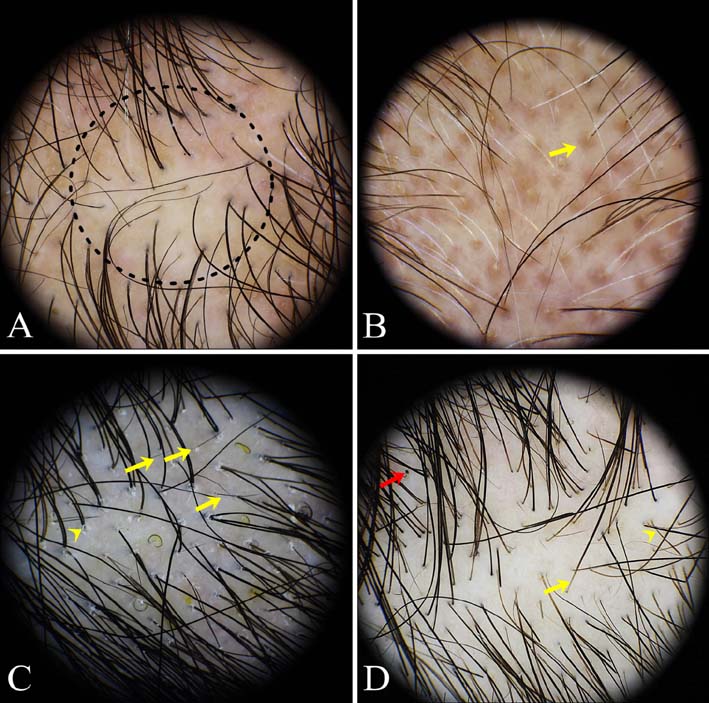
Fig. 3 Trichoscopic findings of diffuse hair loss. (A) Androgenetic alopecia: hair diameter diversity (dotted circle). (B) Androgenetic alopecia: peripilar sign (yellow arrow). (C) Telogen effluvium: short nonvellus hairs (yellow arrows), perifollicular scales (yellow arrowhead). (D) Alopecia areata incognita: proximal tapering hairs (yellow arrow), exclamation mark hairs (yellow arrowhead), black dots (red arrow).
Reference
-
1. Ross EK, Vincenzi C, Tosti A. Videodermoscopy in the evaluation of hair and scalp disorders. J Am Acad Dermatol. 2006; 55:799–806.
Article2. Toncić RJ, Lipozencić J, Pastar Z. Videodermoscopy in the evaluation of hair and scalp disorders. Acta Dermatovenerol Croat. 2007; 15:116–118.3. Lacarrubba F, Dall'Oglio F, Rita Nasca M, Micali G. Videodermatoscopy enhances diagnostic capability in some forms of hair loss. Am J Clin Dermatol. 2004; 5:205–208.
Article4. Rudnicka L, Olszewska M, Rakowska A, Slowinska M. Trichoscopy update 2011. J Dermatol Case Rep. 2011; 5:82–88.
Article5. Rudnicka L, Rakowska A, Olszewska M. Trichoscopy: how it may help the clinician. Dermatol Clin. 2013; 31:29–41.6. Miteva M, Tosti A. Hair and scalp dermatoscopy. J Am Acad Dermatol. 2012; 67:1040–1048.
Article7. Tosti A, Torres F. Dermoscopy in the diagnosis of hair and scalp disorders. Actas Dermosifiliogr. 2009; 100:Suppl 1. 114–119.
Article8. Tosti A, Duque-Estrada B. Dermoscopy in hair disorders. J Egypt Women Dermatol Soc. 2010; 7:1–4.9. Karadağ Köse Ö, Güleç AT. Clinical evaluation of alopecias using a handheld dermatoscope. J Am Acad Dermatol. 2012; 67:206–214.
Article10. Inui S, Nakajima T, Nakagawa K, Itami S. Clinical significance of dermoscopy in alopecia areata: analysis of 300 cases. Int J Dermatol. 2008; 47:688–693.
Article11. Shim WH, Jwa SW, Song M, Kim HS, Ko HC, Kim BS, et al. Dermoscopic approach to a small round to oval hairless patch on the scalp. Ann Dermatol. 2014; 26:214–220.
Article12. Fu JM, Starace M, Tosti A. A new dermoscopic finding in healthy children. Arch Dermatol. 2009; 145:596–597.
Article13. Abraham LS, Torres FN, Azulay-Abulafia L. Dermoscopic clues to distinguish trichotillomania from patchy alopecia areata. An Bras Dermatol. 2010; 85:723–726.14. Rakowska A, Slowinska M, Olszewska M, Rudnicka L. New trichoscopy findings in trichotillomania: flame hairs, V-sign, hook hairs, hair powder, tulip hairs. Acta Derm Venereol. 2014; 94:303–306.
Article15. Tangjaturonrusamee C, Piraccini BM, Vincenzi C, Starace M, Tosti A. Tinea capitis mimicking folliculitis decalvans. Mycoses. 2011; 54:87–88.
Article16. Slowinska M, Rudnicka L, Schwartz RA, Kowalska-Oledzka E, Rakowska A, Sicinska J, et al. Comma hairs: a dermatoscopic marker for tinea capitis: a rapid diagnostic method. J Am Acad Dermatol. 2008; 59:5 Suppl. S77–S79.17. Lacarrubba F, Verzì AE, Micali G. Newly described features resulting from high-magnification dermoscopy of tinea capitis. JAMA Dermatol. 2015; 151:308–310.
Article18. Kossard S, Zagarella S. Spotted cicatricial alopecia in dark skin. A dermoscopic clue to fibrous tracts. Australas J Dermatol. 1993; 34:49–51.19. Deloche C, de Lacharrière O, Misciali C, Piraccini BM, Vincenzi C, Bastien P, et al. Histological features of peripilar signs associated with androgenetic alopecia. Arch Dermatol Res. 2004; 295:422–428.
Article20. Rakowska A, Slowinska M, Kowalska-Oledzka E, Olszewska M, Rudnicka L. Dermoscopy in female androgenic alopecia: method standardization and diagnostic criteria. Int J Trichology. 2009; 1:123–130.
Article21. Inui S, Nakajima T, Itami S. Scalp dermoscopy of androgenetic alopecia in Asian people. J Dermatol. 2009; 36:82–85.
Article22. de Lacharrière O, Deloche C, Misciali C, Piraccini BM, Vincenzi C, Bastien P, et al. Hair diameter diversity: a clinical sign reflecting the follicle miniaturization. Arch Dermatol. 2001; 137:641–646.23. Molina L, Donati A, Valente NS, Romiti R. Alopecia areata incognita. Clinics (Sao Paulo). 2011; 66:513–515.
Article24. Tosti A, Whiting D, Iorizzo M, Pazzaglia M, Misciali C, Vincenzi C, et al. The role of scalp dermoscopy in the diagnosis of alopecia areata incognita. J Am Acad Dermatol. 2008; 59:64–67.
Article25. Werner B, Mulinari-Brenner F. Clinical and histological challenge in the differential diagnosis of diffuse alopecia: female androgenetic alopecia, telogen effluvium and alopecia areata--part II. An Bras Dermatol. 2012; 87:884–890.
Article26. Rudnicka L, Olszewska M, Rakowska A, Czuwara J. Rudnicka L, Olszewska M, Rakowska A, editors. Atlas of trichoscopy. 1st ed. London: Springer-Verlag;2012. p. 206–235.
- Full Text Links
-
- Actions
-
Cited
- CITED
-
- Close
- Share
-
- Similar articles
-
- Representative Trichoscopic Findings of Outpatients with Androgenetic Alopecia and Alopecia Areata
- A Clinical Study of the Normal Values and Guideline for Hair Pull Test in Koreans
- Alopecia
- Hair characteristics and androgenetic alopecia in Koreans
- A Case of Hair Structure Abnormality Associated with Iron Deficiency Anaemia
