Ann Dermatol.
2016 Apr;28(2):273-274. 10.5021/ad.2016.28.2.273.
A Case of Cutaneous Protothecosis in an Immunocompetent Patient
- Affiliations
-
- 1Department of Dermatology, Ewha Womans University School of Medicine, Seoul, Korea. uwon313@ewha.ac.kr
- 2Department of Pathology, Ewha Womans University School of Medicine, Seoul, Korea.
- KMID: 2171398
- DOI: http://doi.org/10.5021/ad.2016.28.2.273
Abstract
- No abstract available.
MeSH Terms
Figure
-
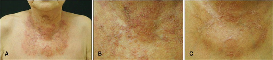
Fig. 1 (A) Several crusted papules on an erythematous telangiectactic patch on the anterior neck and upper chest. (B) Close-up view. (C) Improved skin lesion with fibrotic scar after treatment with oral itraconazole 200 mg/day and topical sertaconazole application for 3 months.
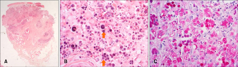
Fig. 2 (A) Granulomatous inflammation with necrosis in the dermis (H&E, ×12.5). (B) Multiple sporangia with in morula-like or cartwheel-like appearance (arrow) (H&E, ×400). (C) Multiple morula-like appearances highlighted by Periodic acid-Schiff stain (×400).
Reference
-
1. Mayorga J, Barba-Gómez JF, Verduzco-Martínez AP, Muñoz-Estrada VF, Welsh O. Protothecosis. Clin Dermatol. 2012; 30:432–436.
Article2. Seok JY, Lee Y, Lee H, Yi SY, Oh HE, Song JS. Human cutaneous protothecosis: report of a case and literature review. Korean J Pathol. 2013; 47:575–578.
Article3. Lass-Flörl C, Mayr A. Human protothecosis. Clin Microbiol Rev. 2007; 20:230–242.
Article4. Hillesheim PB, Bahrami S. Cutaneous protothecosis. Arch Pathol Lab Med. 2011; 135:941–944.
Article5. Todd JR, King JW, Oberle A, Matsumoto T, Odaka Y, Fowler M, et al. Protothecosis: report of a case with 20-year follow-up, and review of previously published cases. Med Mycol. 2012; 50:673–689.
Article
- Full Text Links
-
- Actions
-
Cited
- CITED
-
- Close
- Share
-
- Similar articles
-
- A Case of Protothecosis on Scalp and Face in the Immunocompetent Patient
- A Case of Cutaneous Protothecosis
- Human Cutaneous Protothecosis: Report of a Case and Literature Review
- A Case of Cutaneous Protothecosis
- Two Cases of Cutaneous Protothecosis : Unique Histopathological Findings with Crystal Violet Staining and the Therapeutic Effect of Itraconazole
