Anat Cell Biol.
2014 Mar;47(1):40-43. 10.5115/acb.2014.47.1.40.
Normal stress pattern of the pubic symphysis
- Affiliations
-
- 1Department of Anatomy, Faculty of Medicine, Dokuz Eylul University, Izmir, Turkey. cigdem.icke@deu.edu.tr
- 2Department of Anatomy, University of Cologne, Cologne, Germany.
- KMID: 2168855
- DOI: http://doi.org/10.5115/acb.2014.47.1.40
Abstract
- The pelvic ring is stressed by external forces: by partial body weight, by ligament tension, and by muscles forces stabilizing the hip joints. For the symphysis ossis pubis there exist data concerning the type and magnitude of stresses. In one-leg-standing pressure, shear forces are predominant, and in both-leg-standing tensile forces are acting on the pelvic ring. Rupture of the symphysis is problematic due to the variety of its movements. Most literature descriptions of stress in the symphysis reflect only the frontal plane. Our intention was to make morphological as well as experimental investigations on the symphysis ossis pubis to delineate how it will be stressed in the horizontal plane. Twenty pubic bones taken from embalmed adult human cadavers (12 male, 8 female) were used. Horizontal and frontal slices (3 mm thick) of the symphyseal part of the os pubis were made. X-rays and densitometric analysis were performed. The width of the symphysis cartilage in the dorsal and the ventral regions was measured on 15 whole skeleton specimens coming from adult human cadavers. For experimental study an embalmed pelvic ring which had no abnormality was used. The symphysis pubis was cut completely in the midsagittal plane and then the ring was stressed via the cranial sacrum. Our results demonstrate that the symphysis is stressed by bending in the horizontal plane in one-leg-standing. In both-leg-standing the symphysis is stressed by tensile forces.
Keyword
MeSH Terms
Figure
-

Fig. 1 Study experimental design.
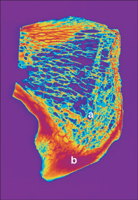
Fig. 2 Arrangement of bone trabeculae in the frontal plane. a, run rectangularly to the compact bone delineating the symphyseal cleft; b, compact bone lamella is thicker and denser in the caudal part.
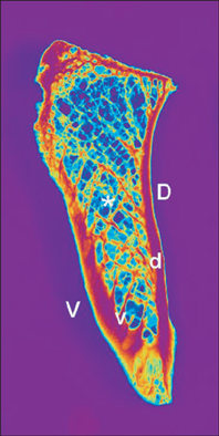
Fig. 3 Arrangement of the bone trabeculae in the horizontal plane. d, dorsal bundle; v, ventral bundle; D, dorsal cortical bone; V, ventral cortical bone. The D was thicker and more dense than the V. *Bone trabeculae bundles intersecting each other.
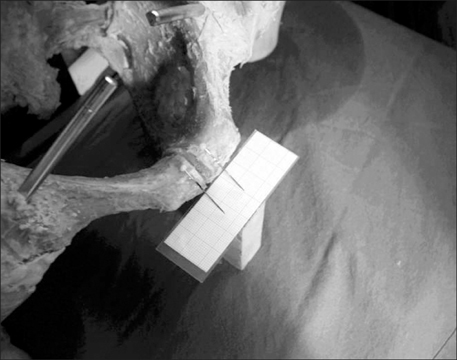
Fig. 4 In one-leg-standing the symphyseal cleft is narrowest in its caudal part.
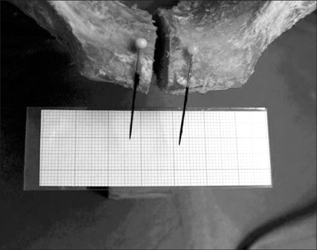
Fig. 5 In both-leg-standing the symphyseal cleft widens in all its parts equally.
Reference
-
1. Ries M, Pugh J, Au JC, Gurtowski J, Dee R. Normal pelvic strain pattern in vitro. J Biomed Eng. 1989; 11:398–402.2. Omar IM, Zoga AC, Kavanagh EC, Koulouris G, Bergin D, Gopez AG, Morrison WB, Meyers WC. Athletic pubalgia and "sports hernia": optimal MR imaging technique and findings. Radiographics. 2008; 28:1415–1438.3. Walheim G, Olerud S, Ribbe T. Mobility of the pubic symphysis. Measurements by an electromechanical method. Acta Orthop Scand. 1984; 55:203–208.4. Dalstra M, Huiskes R, Odgaard A, van Erning L. Mechanical and textural properties of pelvic trabecular bone. J Biomech. 1993; 26:523–535.5. Fabeck L, Descamps PY, Bourgois R, Dhem A. Contribution to the study of pelvic stress during weight-bearing. Role of the pubic branch and trabecular bone. Rev Chir Orthop Reparatrice Appar Mot. 1994; 80:181–187.6. MacAvoy MC, McClellan RT, Goodman SB, Chien CR, Allen WA, van der Meulen MC. Stability of open-book pelvic fractures using a new biomechanical model of single-limb stance. J Orthop Trauma. 1997; 11:590–593.7. Vrahas M, Hern TC, Diangelo D, Kellam J, Tile M. Ligamentous contributions to pelvic stability. Orthopedics. 1995; 18:271–274.8. Pauwels F. Gesammelte abhandlungen zur funktionellen anatomie des bewegungsapparates. Berlin: Springer-Verlag;1965. p. 183–196.9. Holm NJ. The internal stress pattern of the os coxae. Acta Orthop Scand. 1980; 51:421–428.10. Sciascia R. Anatomic and radiologic observations on the general architecture of the spongy substance of the human os coxae. Ann Ital Chir. 1967; 43:1087–1109.11. Euler E. Das Becken: Anatomie, Biomechanik, Frakturver sorgung und Tumorprothetik. Berlin: Springer-Verlag;1966. p. 3–33.12. Dalstra M, Huiskes R. Load transfer across the pelvic bone. J Biomech. 1995; 28:715–724.13. Goel VK, Valliappan S, Svensson NL. Stresses in the normal pelvis. Comput Biol Med. 1978; 8:91–104.14. Goel VK, Svensson NL. Forces on the pelvis. J Biomech. 1977; 10:195–200.15. Varga E, Hearn T, Powell J, Tile M. Effects of method of internal fixation of symphyseal disruptions on stability of the pelvic ring. Injury. 1995; 26:75–80.16. Meissner A, Fell M, Wilk R, Boenick U, Rahmanzadeh R. Biomechanical investigation of the pubic symphysis. Which forces induce mobility of the symphysis under physiological conditions? Unfallchirurg. 1996; 99:415–421.
- Full Text Links
-
- Actions
-
Cited
- CITED
-
- Close
- Share
-
- Similar articles
-
- Outcome of Internal Fixation and Corticocancellous Grafting of Symphysis Pubis Diastasis Which Developed after Malunion of Pubic Rami Fracture
- Separation of the Symphysis Pubis: Report of 5 Cases
- Unusual Dislocation of the Symphysis Pubis: A Case Report
- Stress Fractures in Pubic Bone: Report of 3 Cases
- Role of Transvaginal Ultrasonography in Stress Urinary Incontinence
