Fetal development of the minor lung segment
- Affiliations
-
- 1Department of Anatomy, Tokyo Dental College, Tokyo, Japan. noguchitaku@tdc.ac.jp
- 2Division of Internal Medicine, Iwamizawa Kojin-kai Hospital, Iwamizawa, Japan.
- 3Department of Anatomy and Embryology II, Faculty of Medicine, Universidad Complutense, Madrid, Spain.
- KMID: 2168852
- DOI: http://doi.org/10.5115/acb.2014.47.1.12
Abstract
- The mediobasal segment (S7) of the right lung has been considered to correspond to the cardiac lobe generally seen in mammals. To investigate fetal development of the right mediobasal segmental bronchus (B7), we examined paraffin-embedded serial sections of 15 embrynic and fetal lungs at 7-8 weeks (serial sections) as well as semiserial sections of 8 fetuses at 15-18 weeks (semiserial sections). All of the smaller specimens did not contain B7, but 2 of the 8 larger specimens carried B7: one was found in the immediately anterior side of the inferior pulmonary vein, while in the other, the subdivisions (B7a, B7b) were overriding the vein. Although the incidence might be underestimated because of observations using semiserial sections, the B7 was most likely to develop secondarily during a period from 8 to 15 weeks. Fetal topographical changes (mainly, the descent) of the middle lobe and the inferior pulmonary vein might relate with the secondarily budding of B7. The present result does not reduce a clinical relevance of B7 as a segmental bronchus of the lung segment system.
Figure
-
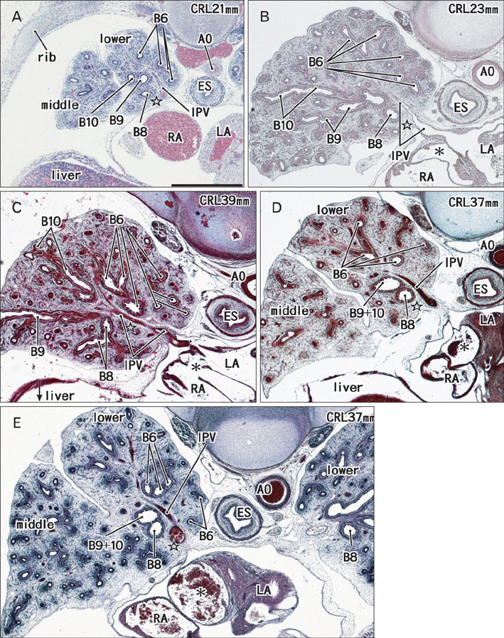
Fig. 1 Five right lung specimens at 7-8 weeks. Panels are shown in order from the smaller lung to the larger one. Note that a specimen with 39 mm crown-rump length (CRL) carries a lung smaller than those in specimens with 37 mm CRL. Panels (A), (D), and (E) include the middle and lower lobes, while panels (B) and (C) the lower lobe only. Star indicates an area in which the segmental bronchus VII is likely to present in the immediately anterior side of the inferior pulmonary vein (IPV). Asterisk indicates a cavity through the oval window between the right atrium (RA) and left atrium (LA). AO, aorta; B6, B8, B9, B10, segmental bronchi VI, VIII, IX, and X; B9+10, a common trunk bronchus with the segmental bronchi IX and X; ES, esophagus. All panels are prepared at the same magnification. Scale bar in panel (A)=1 mm (A-E).
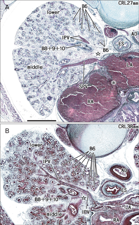
Fig. 2 Two right lung specimens at 7-8 weeks. Note that a specimen with 27 mm crown-rump length (CRL) (A) carries a lung larger than those in specimens with 37 or 39 mm CRL shown in Fig. 1. Both panels include the middle and lower lobes. Star indicates an area in which the segmental bronchus VII is likely to present in the immediately anterior side of the inferior pulmonary vein (IPV). Asterisk indicates a cavity through the oval window between the right atrium (RA) and left atrium (LA). AO, aorta; B6, segmental bronchus VI; B8+9+10, a common trunk bronchus with the segmental bronchi VIII, IX, and X; ES, esophagus; SPV, superior pulmonary vein. The two panels are prepared at the same magnification. Scale bar in panel (A)=1 mm (A, B).
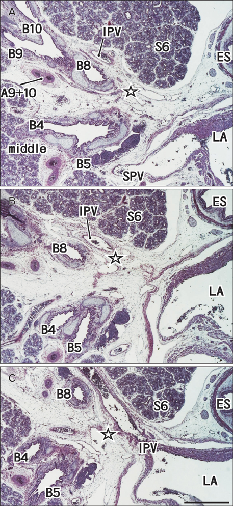
Fig. 3 A 16-week specimen with segmental bronchus VII. Panel (A) (or C) is the most superior (or inferior) in the figure. Intervals between panels are 0.3 mm (A-B, B-C). Star indicates an area in which the segmental bronchus VII is likely to present in the immediately anterior side of the inferior pulmonary vein (IPV). A9+10, a common trunk artery of the segmental arteries IX and X; B4, B5, B6, B8, B9, and B10, segmental bronchi IV, V, VI, VIII, IX, and X; ES, esophagus; LA, left atrium; S6, segment VI; SPV, superior pulmonary vein. All panels are prepared at the same magnification and include the middle and lower lobes. Scale bar in panel (C)=1 mm (A-C).
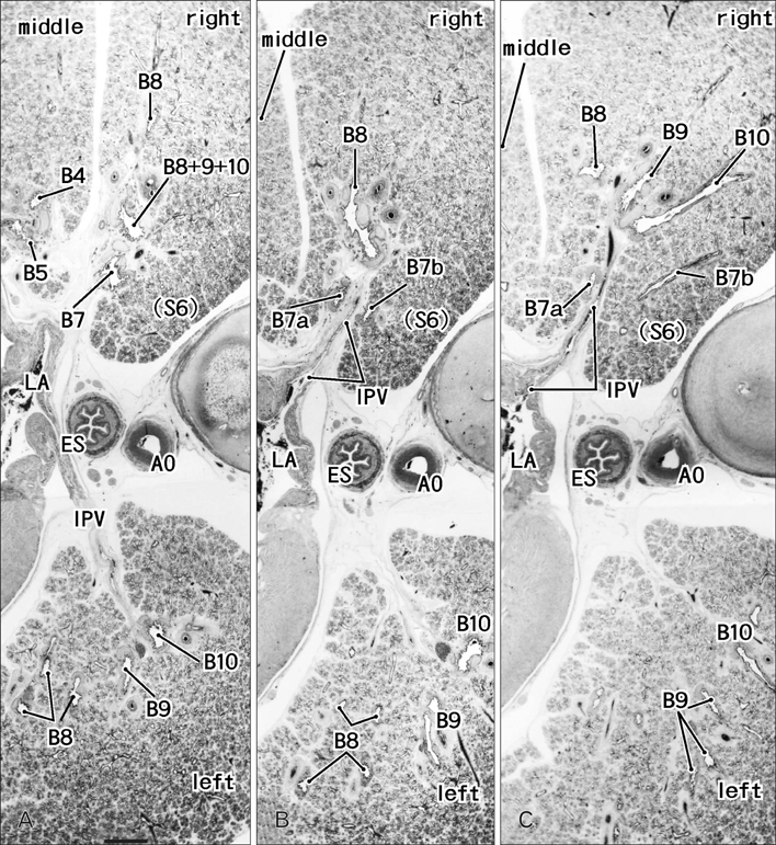
Fig. 4 An 18-week specimen without segmental bronchus VII. Panel (A) (or C) is the most superior (or inferior) in the figure. Intervals between panels are 0.5 mm (A-B, B-C). The topographical orientation (posterior, the right-hand side of each panel) is different from Figs. 1, 2, 3. Segmental bronchus VII (B7) is originated in the 0.5 mm superior side of panel (A), runs inferiorly (A), divides into two subsegmental bronchi (B7a and B8b) in the immediately superior side of the inferior pulmonary vein (IPV in panel B). The B7a (anterior) runs further inferiorly, while the B7b (posterior) runs posterolaterally along the inferolateral margin of the segment VI (S6 in panel C). AO, aorta; B4, B5, B6, B8, B9, and B10, segmental bronchi IV, V, VI, VIII, IX, and X; ES, esophagus; LA, left atrium. All panels are prepared at the same magnification and include the middle and lower lobes. Scale bar in panel (A)=1 mm (A-C).
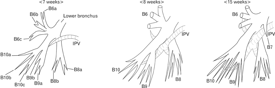
Fig. 5 Schematic representation of the present results. At 7 weeks, except for B7, the bronchial tree in the right lower lobe has already developed at the subsegmental level: B6a, B6b, B6c, B8a, B8b, B9a, B9b, B10a, B10b, B10c. However, the inferior pulmonary vein (IPV) is thin and difficult to identify in the segmental level. At 8 weeks, the vein reaches each of the segments. At 15 weeks, in some specimens, B7 is seen crossing the anterior side of the vein.
Reference
-
1. Williams PL. Gray's anatomy. 38th ed. Edinburgh: Churchill Livingstone;1995.2. Yamashita H. Roentgenologic anatomy of the lung. Tokyo: Igaku-Shoin;1978.3. Arai T, Shiozawa M. Pulmonary resections: regional anatomy and surgical procedures. 2nd ed. Tokyo: Asakura Publishing;1992.4. Huntington GS. A critique of the theories of pulmonary evolution in the mammalia. Am J Anat. 1920; 27:99–201.5. Iino T. Cast anatomy of the bronchial tree and ramification patterns of the pulmonary artery and vein in raccoon dogs. Ann Dep Anat Jikei Univ Sch Med. 1962; 24:21–27.6. Iwasaki M, Matsunaga M. Cast anatomy of the bronchial tree and ramification patterns of the pulmonary artery and vein in cats. Ann Dep Anat Jikei Univ Sch Med. 1962; 24:1–15.7. Kijima T. Cast anatomy of the bronchial tree and ramification patterns of the pulmonary artery and vein in raccoon dogs. Ann Dep Anat Jikei Univ Sch Med. 1962; 24:128–142.8. Moriya M. Cast anatomy of the bronchial tree and ramification patterns of the pulmonary artery and vein in goats. Ann Dep Anat Jikei Univ Sch Med. 1962; 24:31–64.9. Nakakuki S. Comparative anatomical studies on the mammalian lung. Bull Fac Agric Tokyo Univ Agric Technol. 1980; 21:1–74.10. Appleton AB. Segments and blood-vessels of the lungs. Lancet. 1944; 244:592–594.11. Boyden EA. Segmental anatomy of the lung. New York: McGraw-Hill;1955.12. Jackson CL, Huber JF. Correlated applied anatomy of the bronchial tree and lungs with a system of nomenclature. Dis Chest. 1943; 9:319–326.13. Bucher U, Reid L. Development of the intrasegmental bronchial tree: the pattern of branching and development of cartilage at various stages of intra-uterine life. Thorax. 1961; 16:207–218.14. Burri PH. Fetal and postnatal development of the lung. Annu Rev Physiol. 1984; 46:617–628.15. DiFiore JW, Wilson JM. Lung development. Semin Pediatr Surg. 1994; 3:221–232.16. Jeffrey PK. The development of large and small airways. Am J Respir Crit Care Med. 1998; 157(5 Pt 2):S174–S180.17. Laudy JA, Wladimiroff JW. The fetal lung. 1: developmental aspects. Ultrasound Obstet Gynecol. 2000; 16:284–290.18. Szpinda M. The normal growth of the pulmonary trunk in human foetuses. Folia Morphol (Warsz). 2007; 66:126–130.19. Mawatari T, Murakami G, Koshino T, Morishita K, Abe T. Posterior pulmonary lobe: segmental and vascular anatomy in human specimens. Clin Anat. 2000; 13:257–262.20. Koshino T, Murakami G, Sato TJ, Tsugane MH, Fujisawa Y, Mawatari T, Abe T. Configurations of the segmental and subsegmental bronchi and arteries in the right upper lobe of the human lung with special reference to their concomitant relations and double subsegmental arterial supply. Anat Sci Int. 2002; 77:64–73.
