Lipomatosis: a diverse form of hemifacial hyperplasia
- Affiliations
-
- 1Department of Oral Medicine, Diagnosis and Radiology, Sri Guru Ram Das Institute of Dental Sciences and Research, Amritsar, India. dr.preets@gmail.com
- 2Department of Oral Medicine, Diagnosis and Radiology, Government Dental College and Hospital, Mumbai, India.
- 3Department of Prosthodontics, Sri Guru Ram Das Institute of Dental Sciences and Research, Amritsar, India.
- 4Department of Oral Medicine, Diagnosis and Radiology, Dr. D.Y. Patil Dental College and Hospital, Navi Mumbai, India.
- KMID: 2167438
- DOI: http://doi.org/10.5624/isd.2012.42.3.191
Abstract
- A case of hemifacial hyperplasia that presented with muscular, skeletal, and dental hyperplasia along with lipomatous infiltration was described. Advanced imaging was useful in identifying the lipomatous infiltration present in the lesion, which raises the possibility of lipomatosis having a diverse presentation in hemifacial hyperplasia. As there was a scarcity of related literature in the field of dentomaxillofacial radiology, this report would make us familiar with its computed tomographic and magnetic resonance image findings.
MeSH Terms
Figure
-
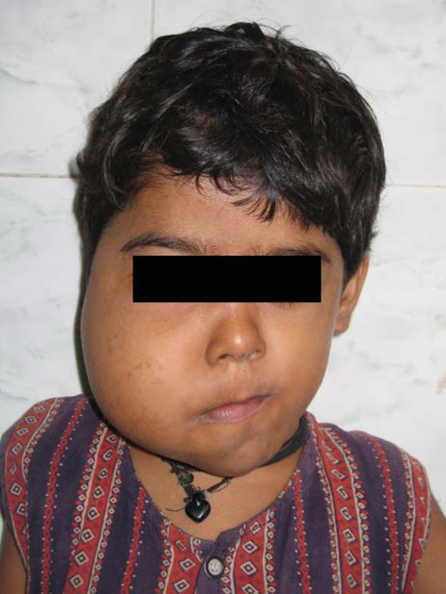
Fig. 1 Facial photograph shows the asymmetrical enlargement of the right side of the face.
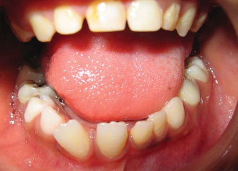
Fig. 2 Intraoral photograph shows the precocious eruption of teeth and enlarged tongue papillae on the right side.
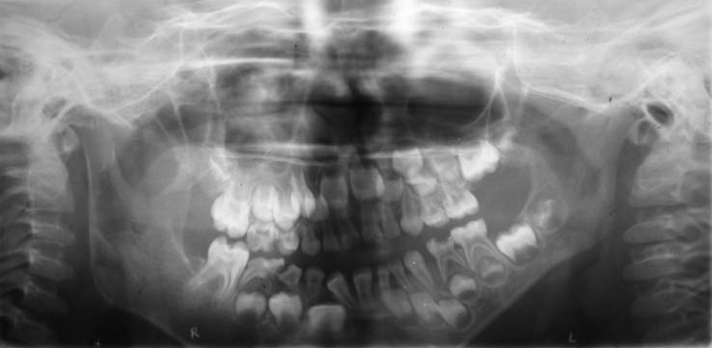
Fig. 3 Panoramic radiograph shows the premature eruption and accelerated root formation of teeth on the right side.
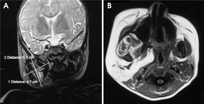
Fig. 4 Coronal (A) and axial (B) MR images show a well-encapsulated mass which appears bright on a T1 weighted image.

Fig. 5 Coronal (A) and axial (B) CT images (bone window and soft tissue window) show the enlargement of the pterygoid plates on the right side and the enlargement on the right side.
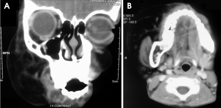
Fig. 6 Contrast enhanced coronal (A) and axial (B) CT images show a well-defined lesion with enhancing septae.
Reference
-
1. Ringrose RE, Jabbour JT, Keele DK. Hemihypertrophy. Pediatrics. 1965. 36:434–448.2. Pollock RA, Newman MH, Burdi AR, Condit DP. Congenital hemifacial hyperplasia: an embryologic hypothesis and case report. Cleft Palate J. 1985. 22:173–184.3. Gessel A. A further study of the nature of hemihypertrophy with report of a new case. Am J Med Sci. 1927. 173:542–554.
Article4. Sugiyama M, Tanaka E, Ogawa I, Ishibashi R, Naito K, Ishikawa T. Magnetic resonance imaging in hemifacial hyperplasia. Dentomaxillofac Radiol. 2001. 30:235–238.
Article5. Bou-Haidar P, Taub P, Som P. Hemifacial lipomatosis, a possible subtype of partial hemifacial hyperplasia: CT and MR imaging findings. AJNR Am J Neuroradiol. 2010. 31:891–893.6. De Rosa G, Cozzolino A, Guarino M, Giardino C. Congenital infiltrating lipomatosis of the face: report of cases and review of the literature. J Oral Maxillofac Surg. 1987. 45:879–883.7. Rowe NH. Hemifacial hypertrophy. Review of literature and addition of four cases. Oral Surg Oral Med Oral Pathol. 1962. 15:572–587.
Article8. Azevedo RA, Souza VF, Sarmento VA, Santos JN. Hemifacial hyperplasia: a case report. Quintessence Int. 2005. 36:483–486.
Article9. Fraumeni JF Jr, Geiser CF, Manning MD. Wilm's tumor and congenital hemihypertrophy: report of five new cases and review of literature. Pediatrics. 1967. 40:886–899.
Article10. Rudolph CE, Norvold RW. Congenital partial-hemihypertrophy involving marked malocclusion. J Dent Res. 1944. 23:133–139.
Article11. Yoshimoto H, Yano H, Kobayashi K, Hirano A, Motomura K, Ohtsuru A, et al. Increased proliferative activity of osteoblasts in congenital hemifacial hypertrophy. Plast Reconstr Surg. 1998. 102:1605–1610.12. Rushton MA. A dental abnormality of size and rate. Proc R Soc Med. 1948. 41:490–496.13. Gorlin RJ, Meskin LH. Congenital hemihypertrophy. Review of the literature and report of a case with special emphasis on oral manifestations. J Pediatr. 1962. 61:870–879.
Article14. Islam MN, Bhattacharya I, Ojha J, Bober K, Cohen DM, Green JG. Comparison between true and partial hemifacial hypertrophy. Oral Surg Oral Med Oral Pathol Oral Radiol Endod. 2007. 104:501–509.
Article
- Full Text Links
-
- Actions
-
Cited
- CITED
-
- Close
- Share
-
- Similar articles
-
- Two Cases of Familial Multiple Lipomatosis
- Idiopathic Spinal Epidural Lipomatosis
- Replacement Lipomatosis of the Kidney: A Case Report
- A Case of Pelvic Lipomatosis
- Unusual Thymic Hyperplasia Mimicking Lipomatous Tumor in an Eight-Year-Old Boy with Concomitant Pericardial Lipomatosis and Right Facial Hemihypertrophy
