Assessment of endodontically treated teeth by using different radiographic methods: an ex vivo comparison between CBCT and other radiographic techniques
- Affiliations
-
- 1Department of Dentomaxillofacial Radiology, Faculty of Dentistry, Gazi University, Ankara, Turkey.
- 2Department of Dentomaxillofacial Radiology, Faculty of Dentistry, Ankara University, Ankara, Turkey. dtkivo@yahoo.com
- 3Department of Biostatistics, Faculty of Medicine, Ankara University, Ankara, Turkey.
- 4Department of Endodontics, Tepebasi Dental Health Center, Ankara, Turkey.
- KMID: 2167430
- DOI: http://doi.org/10.5624/isd.2012.42.3.129
Abstract
- PURPOSE
To compare different radiographic methods for assessing endodontically treated teeth.
MATERIALS AND METHODS
Root canal treatments were applied in 120 extracted mandibular teeth, which were divided into four groups: (1) ideal root canal treatment (60 teeth), (2) insufficient lateral condensation (20 teeth), (3) root canals filled short of the apex (20 teeth), (4) overfilled root canal treatment (20 teeth). The teeth were imaged using intraoral film, panoramic film, digital intraoral systems (CCD and PSP), CCD obtained with portable X-ray source, digital panoramic, and CBCT images obtained at 0.3 mm3 and 0.2 mm3 voxel size. Images were evaluated separately by three observers, twice. Kappa coefficients were calculated. The percentage of correct readings obtained from each modality was calculated and compared using a t-test (p<0.05).
RESULTS
The intra-observer kappa for each observer ranged between 0.327 and 0.849. The inter-observer kappa for each observer for both readings ranged between 0.312 and 0.749. For the ideal root canal treatment group, CBCT with 0.2 mm3 voxel images revealed the best results. For insufficient lateral condensation, the best readings were found with periapical film followed by CCD and PSP. The assessment of teeth with root canals filled short of the apex showed the highest percentage of correct readings by CBCT and CCD. For the overfilled canal treatment group, PSP images and conventional periapical film radiographs had the best scores.
CONCLUSION
CBCT was found to be successful in the assessment of teeth with ideal root canal treatment and teeth with canals filled short of the apex.
MeSH Terms
Figure
-
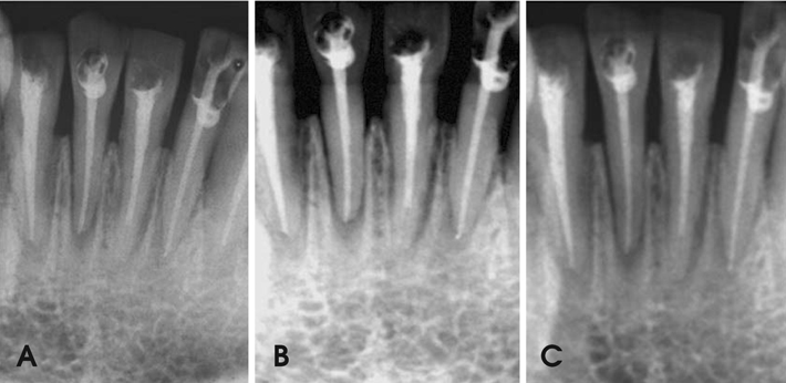
Fig. 1 The images show the different root canal treatments obtained from intraoral techniques. A. Conventional periapical film (Kodak E Speed), B. CCD sensor (Dr. Suni), C. PSP sensor (Digora). From the left: right mandibular lateral incisor with ideal root canal treatment, right mandibular central incisor with insufficient condensation, left mandibular central incisor tooth with root canals filled short of the apex and left mandibular lateral incisor with overfilled root canal treatment.

Fig. 2 Conventional panoramic film (A) and digital panoramic image (B) show the same group of teeth shown in Fig. 1.
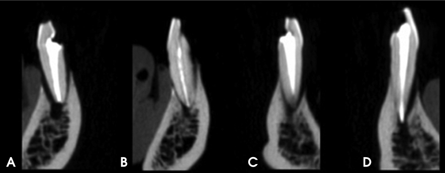
Fig. 3 Images obtained with CBCT unit reconstructed by 0.2mm3 voxels of the teeth shown in Figure 1. A. Ideal root canal treatment. B. Insufficient condensation. C. Root canals filled short of the apex. D. Overfilled root canal treatment.
Reference
-
1. Santos SM, Soares JA, Costa GM, Brito-Júnior M, Moreira AN, de Magalhães CS. Radiographic parameters of quality of root canal fillings and periapical status: a retrospective cohort study. J Endod. 2010. 36:1932–1937.
Article2. Hülsmann M, Peters OA, Dummer PM. Mechanical preparation of root canals: shaping goals, techniques and means. Endod Topics. 2005. 10:30–76.3. Sundqvist G, Figdor D, Persson S, Sjögren U. Microbiologic analysis of teeth with failed endodontic treatment and the outcome of conservative re-treatment. Oral Surg Oral Med Oral Pathol Oral Radiol Endod. 1998. 85:86–93.
Article4. Delivanis PD, Mattison GD, Mendel RW. The survivability of F43 strain of Streptococcus sanguis in root canals filled with guta-percha and Procosol cement. J Endod. 1983. 9:407–410.5. Schilder H. Filling root canals in three dimensions. J Endod. 2006. 32:281–290.
Article6. Shearer AC, Horner K, Wilson NH. Radiovisiography for length estimation in root canal treatment: an in-vitro comparison with conventional radiography. Int Endod J. 1991. 24:233–239.7. Kersten HW, Wesselink PR, Thoden van Velzen SK. The diagnostic reliability of the buccal radiograph after root canal filling. Int Endod J. 1987. 20:20–24.
Article8. Tsesis I, Kamburoğlu K, Katz A, Tamse A, Kaffe I, Kfir A. Comparison of digital with conventional radiography in detection of vertical root fractures in endodontically treated maxillary premolars: an ex vivo study. Oral Surg Oral Med Oral Pathol Oral Radiol Endod. 2008. 106:124–128.
Article9. Kamburoğlu K, Barenboim SF, Kaffe I. Comparison of conventional film with different digital and digitally filtered images in the detection of simulated internal resorption cavities-an ex vivo study in human cadaver jaws. Oral Surg Oral Med Oral Pathol Oral Radiol Endod. 2008. 105:790–797.10. Kamburoğlu K, Tsesis I, Kfir A, Kaffe I. Diagnosis of artificially induced external root resorption using conventional intraoral film radiography, CCD, and PSP: an ex vivo study. Oral Surg Oral Med Oral Pathol Oral Radiol Endod. 2008. 106:885–891.
Article11. Pittayapat P, Oliveira-Santos C, Thevissen P, Michielsen K, Bergans N, Willems G, et al. Image quality assessment and medical physics evaluation of different portable dental X-ray units. Forensic Sci Int. 2010. 201:112–117.
Article12. Pittayapat P, Thevissen P, Fieuws S, Jacobs R, Willems G. Forensic oral imaging quality of hand-held dental X-ray devices: comparison of two image receptors and two devices. Forensic Sci Int. 2010. 194:20–27.
Article13. Goren AD, Bonvento M, Biernacki J, Colosi DC. Radiation exposure with the NOMAD portable X-ray system. Dentomaxillofac Radiol. 2008. 37:109–112.14. Ladeira DB, Cruz AD, Almeida SM, Bóscolo FN. Evaluation of the panoramic image formation in different anatomic positions. Braz Dent J. 2010. 21:458–462.
Article15. Noujeim M, Prihoda T, McDavid WD, Ogawa K, Seki K, Okano T, et al. Pre-clinical evaluation of a new dental panoramic radiographic system based on tomosynthesis method. Dentomaxillofac Radiol. 2011. 40:42–46.
Article16. Ogawa K, Langlais RP, McDavid WD, Noujeim M, Seki K, Okano T, et al. Development of a new dental panoramic radiographic system based on a tomosynthesis method. Dentomaxillofac Radiol. 2010. 39:47–53.
Article17. Dawood A, Patel S, Brown J. Cone beam CT in dental practice. Br Dent J. 2009. 207:23–28.
Article18. Patel S. New dimensions in endodontic imaging: Part 2. Cone beam computed tomography. Int Endod J. 2009. 42:463–475.
Article19. Kamburoğlu K, Murat S, Yüksel SP, Cebeci AR, Horasan S. Detection of vertical root fracture using cone-beam computerized tomography: an in vitro assessment. Oral Surg Oral Med Oral Pathol Oral Radiol Endod. 2010. 109:e74–e81.
Article20. Kamburoğlu K, Ilker Cebeci AR, Gröndahl HG. Effectiveness of limited cone-beam computed tomography in the detection of horizontal root fracture. Dent Traumatol. 2009. 25:256–261.
Article21. Parissis N, Angelopoulos C, Mantegari S, Karamanis S, Masood F, Tsirlis A. A comparison of panoramic image quality between a digital radiography storage phosphor system and a film-based system. J Contemp Dent Pract. 2010. 11:E009–E016.22. Molander B. Panoramic radiography in dental diagnostics. Swed Dent J Suppl. 1996. 119:1–26.23. Alexander JB, Andrews JD. A comparison between xeroradiographs and conventional radiographs as an aid in root canal therapy for maxillary molars. Oral Surg Oral Med Oral Pathol. 1989. 67:443–448.
Article24. Ulusu T, Bodur H, Odabaş ME. In vitro comparison of digital and conventional bitewing radiographs for the detection of approximal caries in primary teeth exposed and viewed by a new wireless handheld unit. Dentomaxillofac Radiol. 2010. 39:91–94.25. Lozano A, Forner L, Llena C. In vitro comparison of root-canal measurements with conventional and digital radiology. Int Endod J. 2002. 35:542–550.
Article26. Friedlander LT, Love RM, Chandler NP. A comparison of phosphor-plate digital images with conventional radiographs for the perceived clarity of fine endodontic files and periapical lesions. Oral Surg Oral Med Oral Pathol Oral Radiol Endod. 2002. 93:321–327.
Article27. Baksi BG, Soğur E, Gröndahl HG. LCD and CRT display of storage phosphor plate and limited cone beam computed tomography images for the evaluation of root canal fillings. Clin Oral Investig. 2009. 13:37–42.28. Liedke GS, da Silveira HE, da Silveira HL, Dutra V, de Figueiredo JA. Influence of voxel size in the diagnostic ability of cone beam tomography to evaluate simulated external root resorption. J Endod. 2009. 35:233–235.
Article29. Moura MS, Guedes OA, De Alencar AH, Azevedo BC, Estrela C. Influence of length of root canal obturation on apical periodontitis detected by periapical radiography and cone beam computed tomography. J Endod. 2009. 35:805–809.
Article30. Huybrechts B, Bud M, Bergmans L, Lambrechts P, Jacobs R. Void detection in root fillings using intraoral analogue, intraoral digital and cone beam CT images. Int Endod J. 2009. 42:675–685.
Article31. Soğur E, Baksi BG, Gröndahl HG. Imaging of root canal fillings: a comparison of subjective image quality between limited cone-beam CT, storage phosphor and film radiography. Int Endod J. 2007. 40:179–185.
Article32. Sherrard JF, Rossouw PE, Benson BW, Carrillo R, Buschang PH. Accuracy and reliability of tooth and root lengths measured on cone-beam computed tomographs. Am J Orthod Dentofacial Orthop. 2010. 137:S100–S108.
Article
- Full Text Links
-
- Actions
-
Cited
- CITED
-
- Close
- Share
-
- Similar articles
-
- Retrospective clinical and radiographic evaluation of restored endodontically treated teeth
- Plain Radiography of the Hip: A Review of Radiographic Techniques and Image Features
- Which factors related to apical radiolucency may influence its radiographic detection? A study using CBCT as reference standard
- Correlation between sagittal condylar guidance angles obtained using radiographic and protrusive occlusal record methods
- Persistent pain after successful endodontic treatment in a patient with Wegener’s granulomatosis: a case report
