Intra-abdominal fibromatosis after gastrectomy for gastric cancer
- Affiliations
-
- 1Department of Surgery, Konkuk University Medical Center, Konkuk University School of Medicine, Seoul, Korea. 20090445@kuh.ac.kr
- 2Department of Pathology, Konkuk University Medical Center, Konkuk University School of Medicine, Seoul, Korea.
- KMID: 2167090
- DOI: http://doi.org/10.4174/astr.2014.87.6.331
Abstract
- Intra-abdominal fibromatosis (IAF) may arise either sporadically or in association with familial adenomatous polyposis. The characteristics of fibromatosis are slow-growth, benign histological features, and aggressive local invasion. Surgery remains a reasonable first treatment option. Here, we report 2 cases of a phenomenon rarely described in published literature, IAF after gastrectomy for gastric cancer. Intra-abdominal masses were found during the routine follow-up period in a 50-year-old man who had received a radical subtotal gastrectomy for early gastric cancer. Two mesenteric masses were detected in the upper abdomen by CT and were excised completely along with segments of the jejunum. Another intra-abdominal mass was found in 60-year-old man who had received a radical total gastrectomy for advanced gastric cancer. A 4.2-cm-sized mass was detected in the periumbilical region by follow-up CT and was excised completely along with a segment of the ileum.
Keyword
MeSH Terms
Figure
-
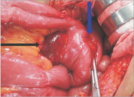
Fig. 1 First tumor in the mesentery of the jejunum near the gastrojejunostomy site (black arrow, mesenteric mass; blue arrow, gastrojejunostomy site).
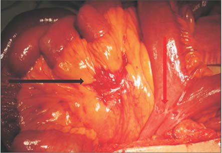
Fig. 2 Second tumor in the mesentery of the jejunum (black arrow, mesenteric mass; red arrow, jejunojejunostomy site).
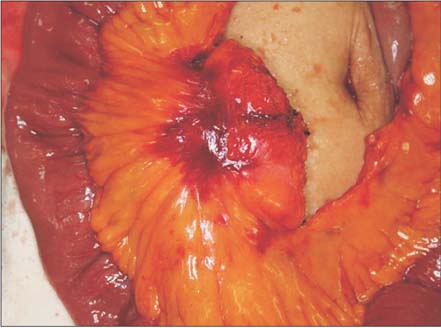
Fig. 3 Second tumor in the mesentery of the jejunum before the jejunum was resected.
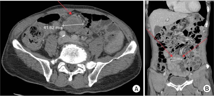
Fig. 4 A 4.2-cm-sized mass in the periumbilical region by CT: (A) horizontal view (red arrow, mesentery mass), (B) coronal view (red arrows, mesentery mass).

Fig. 5 Mild hypermetabolic mass in the periumbilical region by PET/CT (red arrow, mesenteric mass).

Fig. 6 Mesenteric mass and ileum excised completely. (A) resected specimen. (B) cross-sectional view.
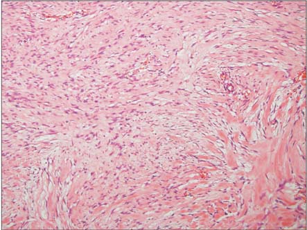
Fig. 7 Histologically, proliferation of spindle-shaped cells with uniform cytologic features and focal dense collagen deposition is seen (H&E, ×100).
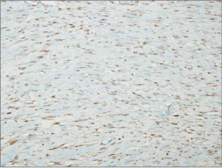
Fig. 8 Immunohistochemically, the tumor cells showed diffuse β-catenin nuclear staining (×200).
Reference
-
1. Lewis JJ, Boland PJ, Leung DH, Woodruff JM, Brennan MF. The enigma of desmoid tumors. Ann Surg. 1999; 229:866–872.2. Komatsu S, Ichikawa D, Kurioka H, Koide K, Ueshima Y, Shioaki Y, et al. Intra-abdominal desmoid tumor mimicking lymph node recurrence after gastrectomy for gastric cancer. J Gastroenterol Hepatol. 2006; 21:1224–1226.3. Clark SK, Phillips RK. Desmoids in familial adenomatous polyposis. Br J Surg. 1996; 83:1494–1504.4. Sturt NJ, Clark SK. Current ideas in desmoid tumours. Fam Cancer. 2006; 5:275–285.5. Zhu H, Chen H, Zhang S, Peng W. Intra-abdominal fibromatosis: differentiation from gastrointestinal stromal tumour based on biphasic contrast-enhanced CT findings. Clin Radiol. 2013; 68:1133–1139.6. Kasper B, Strobel P, Hohenberger P. Desmoid tumors: clinical features and treatment options for advanced disease. Oncologist. 2011; 16:682–693.7. Tolan S, Shanks JH, Loh MY, Taylor B, Wylie JP. Fibromatosis: benign by name but not necessarily by nature. Clin Oncol (R Coll Radiol). 2007; 19:319–326.8. Janinis J, Patriki M, Vini L, Aravantinos G, Whelan JS. The pharmacological treatment of aggressive fibromatosis: a systematic review. Ann Oncol. 2003; 14:181–190.9. Huss S, Nehles J, Binot E, Wardelmann E, Mittler J, Kleine MA, et al. β-catenin (CTNNB1) mutations and clinicopathological features of mesenteric desmoid-type fibromatosis. Histopathology. 2013; 62:294–304.
- Full Text Links
-
- Actions
-
Cited
- CITED
-
- Close
- Share
-
- Similar articles
-
- Mesenteric Fibromatosis Mimicking Recurrence after Distal Gastrectomy for Gastric Cancer
- Abdominal Drainage in the Prevention and Management of Major IntraAbdominal Complications after Total Gastrectomy for Gastric Carcinoma
- Single Port Gastrectomy for Gastric Cancer
- Endoscopic Ultrasound-Guided Transgastric Drainage of an Intra-Abdominal Abscess following Gastrectomy
- Is Laparoscopic Approach Also Safe for the Treatment of Remnant Gastric Cancer?
