Noninvasive monitoring of mouse renal allograft rejection using micro-CT
- Affiliations
-
- 1Department of Urology, Huashan Hospital, Fudan University, Shanghai, China.
- 2Division of Transplantation Immunology, National Research Institute for Child Health and Development, Tokyo, Japan. ri-k@ncchd.go.jp
- 3AIDS Research Center, National Institute of Infectious Diseases, Tokyo, Japan.
- KMID: 2166996
- DOI: http://doi.org/10.4174/astr.2015.88.5.276
Abstract
- PURPOSE
Acute renal graft rejection can only be definitively diagnosed by renal biopsy. However, biopsies carry a risk of renal transplant injury and loss. Micro-CT is widely used in preclinical studies of small animals. Here, we propose micro-CT could noninvasively monitor and evaluate renal location and function in a mouse kidney transplant model.
METHODS
Orthotopic kidney transplantation was performed in a BALB/c -to- C57BL/6j or C57BL/6j-to- C57BL/6j mouse model. After optimizing imaging techniques, five mice were imaged with micro-CT and the findings were verified histologically.
RESULTS
Micro-CT can monitor and evaluate renal location and function after orthotopic kidney transplantation. There were no mice deaths while renal transplants were failure.
CONCLUSION
We propose that graft micro-CT imaging is a new option that is noninvasive and specific, and can aid in early detection and follow-up of acute renal rejection. This method is potentially useful to improve posttransplant rejection monitoring.
Keyword
MeSH Terms
Figure
-
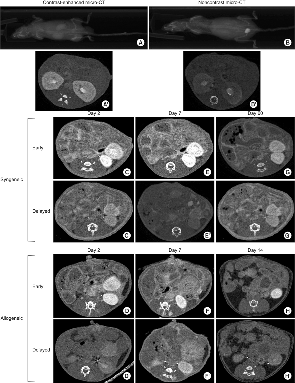
Fig. 1 Imaging of the mouse native and transplanted kidney. The statistical atlas-based registration was validated based on both noncontrast (A/A') and contrast-enhanced (B/B') micro-CT images. Micro-CT images of the transplanted mouse kidneys day 2 (C/C' and D/D'), 7 (E/E' and F/F'), 14 (G/G'), and 60 (H/H') post operation were obtained. Early (5 minutes; without apostrophe: C to H) and delayed (30 minutes: with apostrophe: C' to H') micro-CT scans were taken to confirm the presence of living renal transplant. Living transplanted kidneys were detected by micro-CT in left abdomen at two days after surgery (C/C' and D/D'). In syngeneic transplanted models, the living renal transplant can be detected at more than two months (G/G'). However, allograft transplanted kidneys were found nonfunctioning at day 7 (F/F') and 14 (H/H') post operation.

Fig. 2 Histological assessment. H&E staining was performed on the syngeneic transplanted kidneys at day 7 (A) and 60 (B) and allograft transplanted kidneys at day 7 (C) and 14 (D). Comparison with syngeneic transplanted kidney, the allograft transplanted kidneys showed venulitis, mild tubulitis, interstitial infiltration, and severe tubulitis. Bar indicates 200 µm and Bar in inset indicates 100 µm.
Reference
-
1. Ge F, Gong W. Strategies for successfully establishing a kidney transplant in a mouse model. Exp Clin Transplant. 2011; 9:287–294.2. Tse GH, Hughes J, Marson LP. Systematic review of mouse kidney transplantation. Transpl Int. 2013; 26:1149–1160.3. Schambach SJ, Bag S, Schilling L, Groden C, Brockmann MA. Application of micro-CT in small animal imaging. Methods. 2010; 50:2–13.4. Azuma H, Isaka Y, Nomi H, Inamoto T, Li XK, Hounig T, et al. Induction of donorspecific tolerance using superagonistic CD28 antibody in rat renal allografts: regulatory T-cell expansion before engraftment may be important. Transplantation. 2010; 90:1328–1335.5. Abe T, Li XK, Yazawa K, Hatayama N, Xie L, Sato B, et al. Hydrogen-rich University of Wisconsin solution attenuates renal cold ischemia-reperfusion injury. Transplantation. 2012; 94:14–21.6. Willmann JK, van Bruggen N, Dinkelborg LM, Gambhir SS. Molecular imaging in drug development. Nat Rev Drug Discov. 2008; 7:591–607.7. Soltysiak P, Saxena AK. Micro-computed tomography for implantation site imaging during in situ oesophagus tissue engineering in a live small animal model. J Tissue Eng Regen Med. 2009; 3:573–576.8. Masyuk TV, Radtke BN, Stroope AJ, Banales JM, Masyuk AI, Gradilone SA, et al. Inhibition of Cdc25A suppresses hepato-renal cystogenesis in rodent models of polycystic kidney and liver disease. Gastroenterology. 2012; 142:622–633.e4.9. Vierling JM, Hreha G, Wang H, Braun M. The role of biliary epithelial cells in the immunopathogenesis of non-suppurative destructive cholangitis in murine hepatic graft-versus-host disease. Trans Am Clin Climatol Assoc. 2011; 122:326–335.10. Paulus MJ, Gleason SS, Kennel SJ, Hunsicker PR, Johnson DK. High resolution X-ray computed tomography: an emerging tool for small animal cancer research. Neoplasia. 2000; 2:62–70.11. Marx J. Imaging. Animal models: live and in color. Science. 2003; 302:1880–1882.12. Maehara N. Experimental microcomputed tomography study of the 3D microangioarchitecture of tumors. Eur Radiol. 2003; 13:1559–1565.13. Cai QY, Lee H, Kim EJ, Moon H, Chang K, Rho J, et al. Magnetic resonance imaging of superparamagnetic iron oxide-labeled macrophage infiltrates in acute-phase renal ischemia-reperfusion mouse model. Nanomedicine. 2012; 8:365–373.14. Reuter S, Schnockel U, Schroter R, Schober O, Pavenstadt H, Schafers M, et al. Noninvasive imaging of acute renal allograft rejection in rats using small animal FFDG-PET. PLoS One. 2009; 4:e5296.
- Full Text Links
-
- Actions
-
Cited
- CITED
-
- Close
- Share
-
- Similar articles
-
- Immunologic monitoring in kidney transplant recipients
- Diagnosis of renal transplant rejection: Banff classification and beyond
- Impact of the Pattern of Acute Rejection Episodes on Graft Survival
- Allograft Immune Reaction of Kidney Transplantation: Part 1. Mechanism of Allograft Rejection
- A Case of Spontaneous Rupture of REnal Allograft
