A Case of Synchronous Double Primary Cancer of the Penis and Urinary Bladder
- Affiliations
-
- 1Department of Internal Medicine, Eulji University School of Medicine, Daejeon, Korea. lee982023@eulji.ac.kr
- 2Department of Pathology, Eulji University School of Medicine, Daejeon, Korea.
- 3Department of Radiation Oncology, Eulji University School of Medicine, Daejeon, Korea.
- 4Department of Urology, Eulji University School of Medicine, Daejeon, Korea.
Abstract
- Multiple primary cancers are the occurrence of more than two cancers of different origin in an individual. Penile cancer is a rare disease, and finding it combined with other cancers is even rarer. A 64-year-old man with a painful penile mass was referred to us from a primary urological clinic. We performed a biopsy of the penile mass and the histology revealed a well-differentiated squamous cell carcinoma. Abdominal computed tomography showed a localized bladder tumor with inguinal lymphadenopathy. The patient underwent a partial penectomy, transurethral resection of the bladder tumor and inguinal lymph node dissection. The histology of the bladder tumor was high-grade papillary carcinoma, and that of the lymph node was squamous cell carcinoma. The penile and bladder tumors were in stage II (T1N1M0) and stage I (T1N0M0), respectively. We successfully treated the patient with adjuvant radiotherapy and systemic chemotherapy.
MeSH Terms
Figure
-
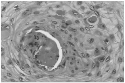
Fig. 1 The pathologic features of the penile tumor (H&E stain, ×200). The microphotograph showed an invasive squamous cell carcinoma with well-differentiated mature squamous cells and abundant keratin pearls and occasional well-developed intercellular bridges.
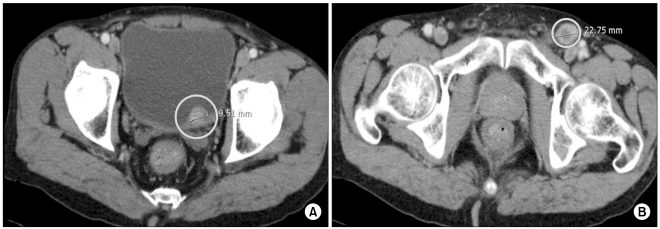
Fig. 2 (A) The abdominal CT showed an approximately 1 cm sized enhancing luminal protruding polypoid mass on the left posterior wall of the urinary bladder. (B) The abdominal CT showed a 2.2 cm enlarged lymph node with a tendency for focal infiltration in the left inguinal area.
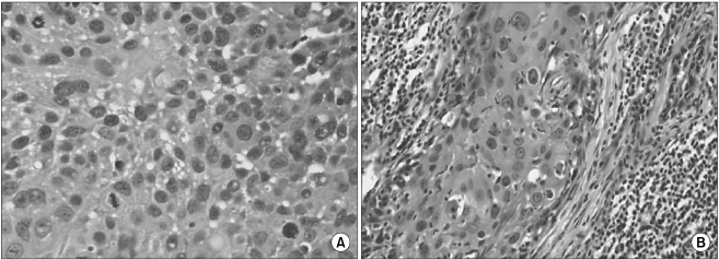
Fig. 3 (A) The pathologic features of the bladder tumor (H&E stain, ×200). The microphotograph showed an invasive urothelial carcinoma with more nuclear pleomorphism, anaplasia and many mitotic figures. (B) The pathologic features of the left inguinal lymph node (H&E stain, ×100). The microphotograph showed a metastatic squamous cell carcinoma with less differentiation and more nuclear pleomorphism and mitotic activity.
Reference
-
1. Billroth T. General surgical pathology and therapeutics in 51 Vorlesungen: a textbook for students and physicians in fifty-one lectures. 1889. 14th ed. Berlin, DE: G. Rerimer.2. Warren S, Gates O. Multiple primary malignant tumors; Surgery of literature and statistical study. Am J Cancer. 1932; 16:1358–1414.3. Wegner HE. Multiple primary cancers in urologic patients: Audit of 19-year experience in Berlin and review of the literature. Urology. 1992; 39:231–236. PMID: 1546416.
Article4. Lee C, Lee ES, Choi H, Koh SK, Lee JM, Chai SE, et al. Incidence estimation of genitourinary cancer in Korea. J Korean Med Sci. 1992; 7:154–161. PMID: 1524728.
Article5. Barnholtz-Sloan JS, Maldonado JL, Pow-sang J, Giuliano AR. Incidence trends in primary malignant penile cancer. Urol Oncol. 2007; 25:361–367. PMID: 17826651.
Article6. Kim WJ, Chung JI, Hong JH, Kim CS, Jung SI, Yoon DK. Epidemiological study for urologic cancer in Korea (1998-2002). Korean J Urol. 2004; 45:1081–1088.7. Kakizaki H, Abe Y, Sugano O, Kato H. A hundred cases of multiple primary neoplasms in association with genitourinary cancer. Nippon Hinyokika Gakkai Zasshi. 1992; 83:1841–1846. PMID: 1479754.
Article8. Kim CK, Chang JW. Multiple primary malignant tumors. J Korean Surg Soc. 1970; 12:63–71.9. Koo DJ, Yun DS, Lee JJ, Park CJ. Clinical analysis of 65 multiple primary cancer cases. J Korean Surg Soc. 1999; 56:137–142.10. Kang HJ, Choi S, Kim JC, Rhew HY. Multiple primary malignant neoplasms in genitourinary tract. Korean J Urol. 1994; 35:1265–1270.11. Moertel CG, Dockerty MB, Baggenstoss AH. Multiple primary malignant neoplasm. II. Tumors of different tissues or organs. Cancer. 1961; 14:231–237. PMID: 13771653.12. Matzkin H, Braf Z. Multiple primary malignant neoplasms in the genitourinary tract: occurrence and etiology. J Urol. 1989; 142:1–12. PMID: 2659818.
Article13. Dillner J, von Krogh G, Horenblas S, Meijer CJ. Etiology of squamous cell carcinoma of the penis. Scand J Urol Nephrol Suppl. 2000; 205:189–193. PMID: 11144896.
Article
- Full Text Links
-
- Actions
-
Cited
- CITED
-
- Close
- Share
-
- Similar articles
-
- A Case of Metastatic Cancer of the Penis from Urinary Bladder Carcinoma
- A Case of Synchronous Squamous Cell Carcinoma of the Bladder and Transitional Cell Carcinoma of the Ureter
- A Case of Synchronous Double Primary Cancer of Gastric Adenocarcinoma and Thyroid Papillary Carcinoma
- A case report of metachronous triple primary cancers including stomach, bladder and lung
- A case of double primary cancers of uterine endometrium and bladder with unusual histology
