Ann Rehabil Med.
2015 Jun;39(3):488-493. 10.5535/arm.2015.39.3.488.
Computed Tomography as an Objective Measurement Tool for Secondary Lymphedema Treated With Extracorporeal Shock Wave Therapy
- Affiliations
-
- 1Department of Rehabilitation Medicine, Ewha Womans University School of Medicine, Seoul, Korea. acebhs@gmail.com
- KMID: 2165654
- DOI: http://doi.org/10.5535/arm.2015.39.3.488
Abstract
- Two patients with stage three secondary lymphedema of the upper extremities underwent treatment for breast cancer, including surgery, chemotherapy, and radiotherapy. They were examined with computed tomography (CT) before and after extracorporeal shock wave therapy (ESWT). We used a manual tracing method using PiViewSTAR software to calculate the volume of the upper extremities. There was a decrease in the volume of the subcutaneous compartment measured by CT before and after ESWT. CT may be helpful in determining the treatment target area of ESWT and to monitor the effect of treatment by measuring the changes in volume before and after ESWT in patients with lymphedema. Therefore, CT may have good clinical potential for treatment and follow-up in the management of lymphedema.
MeSH Terms
Figure
-

Fig. 1 Comparison of computed tomography of the upper extremity. Before (A) and after (B) treatment in the case 1 patient. Before (C) and after (D) treatment of the case 2 patient.
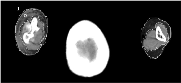
Fig. 2 Computed tomography based on the volume measurement method for lymphedema of the upper extremity whole extremity (1), the sum of bone and muscle (2).
Reference
-
1. Bell RJ, Robinson PJ, Barallon R, Fradkin P, Schwarz M, Davis SR. Lymphedema: experience of a cohort of women with breast cancer followed for four years after diagnosis in Victoria, Australia. Support Care Cancer. 2013; 21:2017–2024. PMID: 23435596.2. Korpan MI, Crevenna R, Fialka-Moser V. Lymphedema: a therapeutic approach in the treatment and rehabilitation of cancer patients. Am J Phys Med Rehabil. 2011; 90(5):Suppl 1. S69–S75. PMID: 21765266.3. Serizawa F, Ito K, Matsubara M, Sato A, Shimokawa H, Satomi S. Extracorporeal shock wave therapy induces therapeutic lymphangiogenesis in a rat model of secondary lymphoedema. Eur J Vasc Endovasc Surg. 2011; 42:254–260. PMID: 21454105.
Article4. Kubo M, Li TS, Kamota T, Ohshima M, Shirasawa B, Hamano K. Extracorporeal shockwave therapy ameliorates secondary lymphedema by promoting lymphangiogenesis. J Vasc Surg. 2010; 52:429–434. PMID: 20670777.5. Bae H, Kim HJ. Clinical outcomes of extracorporeal shock wave therapy in patients with secondary lymphedema: a pilot study. Ann Rehabil Med. 2013; 37:229–234. PMID: 23705118.
Article6. Tinazzi E, Amelio E, Marangoni E, Guerra C, Puccetti A, Codella OM, et al. Effects of shock wave therapy in the skin of patients with progressive systemic sclerosis: a pilot study. Rheumatol Int. 2011; 31:651–656. PMID: 20066427.
Article7. Stanton AW, Badger C, Sitzia J. Non-invasive assessment of the lymphedematous limb. Lymphology. 2000; 33:122–135. PMID: 11019400.8. Roberts CC, Stanton AW, Pullen J, Bull RH, Levick JR, Mortimer PS. Skin microvascular architecture and perfusion studied in human postmastectomy oedema by intravital video-capillaroscopy. Int J Microcirc Clin Exp. 1994; 14:327–334. PMID: 7543461.
Article9. Monnin-Delhom ED, Gallix BP, Achard C, Bruel JM, Janbon C. High resolution unenhanced computed tomography in patients with swollen legs. Lymphology. 2002; 35:121–128. PMID: 12363222.
- Full Text Links
-
- Actions
-
Cited
- CITED
-
- Close
- Share
-
- Similar articles
-
- Clinical Outcomes of Extracorporeal Shock Wave Therapy in Patients With Secondary Lymphedema: A Pilot Study
- Current Concepts in Extracorporeal Shock Wave Therapy
- Treatment of Nonunion of Tibia with Extracorporeal Shock Wave Therapy: A Case Report
- Feasibility of Extracorporeal Shock Wave Therapy for Complex Upper Limb Morbidity in Breast Cancer Patient
- Extracorporeal Shock Wave Therapy for the Treatment of Refractory Plantar Fasciitis
