Ann Rehabil Med.
2015 Dec;39(6):950-956. 10.5535/arm.2015.39.6.950.
Changes in Activation of Abdominal Muscles at Selected Angles During Trunk Exercise by Using Ultrasonography
- Affiliations
-
- 1Department of Physical Medicine and Rehabilitation, Inje University Busan Paik Hospital, Inje University College of Medicine, Busan, Korea. claudjun@naver.com
- KMID: 2165621
- DOI: http://doi.org/10.5535/arm.2015.39.6.950
Abstract
OBJECTIVE
To investigate the changes of activation of the abdominal muscles depending on exercise angles and whether the activation of rectus abdominis differs according to the location, during curl up and leg raise exercises, by measuring the thickness ratio of abdominal muscles using ultrasonography.
METHODS
We examined 30 normal adults without musculoskeletal problems. Muscle thickness was measured in the upper rectus abdominis (URA), lower rectus abdominis (LRA), obliquus externus (EO), obliquus internus (IO), and transversus abdominis (TrA), at pre-determined angles (30degrees, 60degrees, 90degrees) and additionally at the resting angle (0degrees). Muscle thickness ratio was calculated by dividing the resting (0degrees) thickness for each angle, and was used as reflection of muscle activity.
RESULTS
The muscle thickness ratio was significantly different depending on the angles in URA and LRA. For curl up-URA p=0 (30degrees<60degrees), p=0 (60degrees>90degrees), p=0.44 (30degrees<90degrees) and LRA p=0.01 (30degrees<60degrees), p=0 (60degrees>90degrees), p=0.44 (30degrees>90degrees), respectively, by one-way ANOVA test-and for leg raise-URA p=0 (30degrees<60degrees), p=0 (60degrees<90degrees), p=0 (30degrees<90degrees) and LRA p=0.01 (30degrees<60degrees), p=0 (60degrees<90degrees), p=0 (30degrees<90degrees), respectively, by one-way ANOVA test-exercises, but not in the lateral abdominal muscles (EO, IO, and TrA). Also, there was no significant difference in the muscle thickness ratio of URA and LRA during both exercises. In the aspect of muscle activity, there was significant difference in the activation of RA muscle by selected angles, but not according to location during both exercises.
CONCLUSION
According to this study, exercise angle is thought to be an important contributing factor for strengthening of RA muscle; however, both the exercises are thought to have no property of strengthening RA muscle selectively based on the location.
Keyword
Figure
-
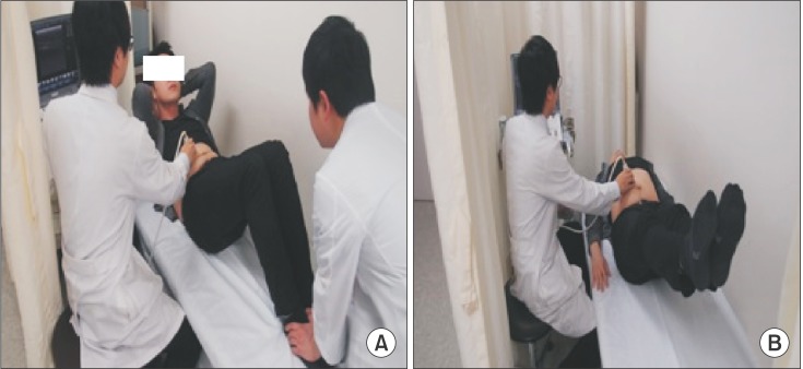
Fig. 1 Images of performing curl up (A) and leg raise (B) exercises while measuring abdominal muscle thickness using ultrasonography.
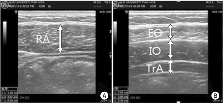
Fig. 2 Ultrasonographic images of rectus (A) and lateral abdominal (B) muscles were made, and thickness was measured from the superficial to deep boundaries of the muscles at the edge of the hypoechoic region. RA, rectus abdominis; EO, obliquus externus; IO, obliquus internus; TrA, transversus abdominis.
Cited by 1 articles
-
Ultrasound Imaging of the Trunk Muscles in Acute Stroke Patients and Relations With Balance Scales
Yunho Kim, Jeeyoung Kim, Heesung Nam, Hyun Dong Kim, Mi Ja Eom, Sang Hoon Jung, Nami Han
Ann Rehabil Med. 2020;44(4):273-283. doi: 10.5535/arm.19125.
Reference
-
1. Kibler WB, Press J, Sciascia A. The role of core stability in athletic function. Sports Med. 2006; 36:189–198. PMID: 16526831.
Article2. Willett GM, Hyde JE, Uhrlaub MB, Wendel CL, Karst GM. Relative activity of abdominal muscles during commonly prescribed strengthening exercises. J Strength Cond Res. 2001; 15:480–485. PMID: 11726260.
Article3. Marchetti PH, Kohn AF, Duarte M. Selective activation of the rectus abdominis muscle during low-intensity and fatiguing tasks. J Sports Sci Med. 2011; 10:322–327. PMID: 24149878.4. Lehman GJ, McGill SM. Quantification of the differences in electromyographic activity magnitude between the upper and lower portions of the rectus abdominis muscle during selected trunk exercises. Phys Ther. 2001; 81:1096–1101. PMID: 11319934.
Article5. Urquhart DM, Hodges PW, Allen TJ, Story IH. Abdominal muscle recruitment during a range of voluntary exercises. Man Ther. 2005; 10:144–153. PMID: 15922235.
Article6. Ishida H, Hirose R, Watanabe S. Comparison of changes in the contraction of the lateral abdominal muscles between the abdominal drawing-in maneuver and breathe held at the maximum expiratory level. Man Ther. 2012; 17:427–431. PMID: 22595657.
Article7. Kim MH, Oh JS. Effects of performing an abdominal hollowing exercise on trunk muscle activity during curl-up exercise on an unstable surface. J Phys Ther Sci. 2015; 27:501–503. PMID: 25729202.
Article8. Yoon TL, Kim KS, Cynn HS. Slow expiration reduces sternocleidomastoid activity and increases transversus abdominis and internal oblique muscle activity during abdominal curl-up. J Electromyogr Kinesiol. 2014; 24:228–232. PMID: 24210796.
Article9. Sarti MA, Monfort M, Fuster MA, Villaplana LA. Muscle activity in upper and lower rectus abdominus during abdominal exercises. Arch Phys Med Rehabil. 1996; 77:1293–1297. PMID: 8976314.
Article10. Duncan M. Muscle activity of the upper and lower rectus abdominis during exercises performed on and off a Swiss ball. J Bodyw Mov Ther. 2009; 13:364–367. PMID: 19761961.
Article11. Clark KM, Holt LE, Sinyard J. Electromyographic comparison of the upper and lower rectus abdominis during abdominal exercises. J Strength Cond Res. 2003; 17:475–483. PMID: 12930172.
Article12. Critchley DJ, Coutts FJ. Abdominal muscle function in chronic low back pain patients: measurement with real-time ultrasound scanning. Physiotherapy. 2002; 88:322–332.13. Rankin G, Stokes M, Newham DJ. Abdominal muscle size and symmetry in normal subjects. Muscle Nerve. 2006; 34:320–326. PMID: 16775833.
Article14. Ferreira PH, Ferreira ML, Nascimento DP, Pinto RZ, Franco MR, Hodges PW. Discriminative and reliability analyses of ultrasound measurement of abdominal muscles recruitment. Man Ther. 2011; 16:463–469. PMID: 21398167.
Article15. Ferreira PH, Ferreira ML, Hodges PW. Changes in recruitment of the abdominal muscles in people with low back pain: ultrasound measurement of muscle activity. Spine (Phila Pa 1976). 2004; 29:2560–2566. PMID: 15543074.16. Stokes IA, Gardner-Morse MG, Henry SM. Abdominal muscle activation increases lumbar spinal stability: analysis of contributions of different muscle groups. Clin Biomech (Bristol, Avon). 2011; 26:797–803.
Article17. Hodges PW, Pengel LH, Herbert RD, Gandevia SC. Measurement of muscle contraction with ultrasound imaging. Muscle Nerve. 2003; 27:682–692. PMID: 12766979.
- Full Text Links
-
- Actions
-
Cited
- CITED
-
- Close
- Share
-
- Similar articles
-
- Effects of the Abdominal Hollowing Technique Applied during Plank Exercises at Different Angles between Ground and the Humerus on Abdominal Stabilization Muscle Activity
- The Effects of Bridge Exercise with One Hip Joint Adduction on Trunk Muscle Thickness
- Muscles Activation of Trunk and Lower-limb during Integrating Bridge Exercise Using Gym Ball in Healthy Individuals
- The Effects of Pilates Mat Exercise on Trunk Muscle Thickness and Balance
- Effects of Squatting with Different Foot Positions on Muscle Activations in Subjects with Genu Varum
