MR Findings of Squamous Cell Carcinoma Arising from Chronic Osteomyelitis of the Tibia: A Case Report
- Affiliations
-
- 1Department of Radiology, Sanggye Paik Hospital, Inje University College of Medicine, Seoul, Korea. merita@paik.ac.kr
- KMID: 2164833
- DOI: http://doi.org/10.3348/jksr.2016.74.5.344
Abstract
- Malignant transformation is a rare and late complication of untreated chronic osteomyelitis. Known radiographic findings of the malignant transformation of chronic osteomyelitis are osteolytic or mixed sclerotic and osteolytic lesions with or without soft tissue mass. But its magnetic resonance (MR) imaging findings are rarely described in the literature. We experienced a case of an 82-year-old man diagnosed with squamous cell carcinoma arising from long standing chronic osteomyelitis of the tibia. Our case indicates that radiologists should consider the possibility of malignant transformation in patients with untreated chronic osteomyelitis, with enhancing soft tissue mass invading and extending through underlying bone cortex and medulla on MR imaging.
MeSH Terms
Figure
-
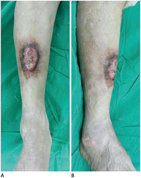
Fig. 1 Anteroposterior (A) and lateral (B) views of the wound. Wound with pus-like discharge is shown on the anterior aspect of the shin. This wound is connected with the medullary cavity of tibia via a draining sinus in the inferior portion of the wound. The wound is accompanied by skin elevation around the wound.
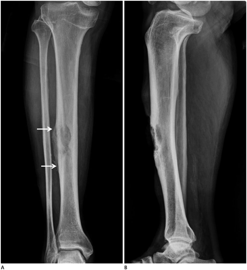
Fig. 2 Simple radiographs of anteroposterior (A) and lateral (B) views of tibia show a partially-defined heterogeneous osteolytic lesion with cortical defect in the anterior aspect of diaphysis of the tibia. Irregular thick solid periosteal reaction is also noted (arrows).
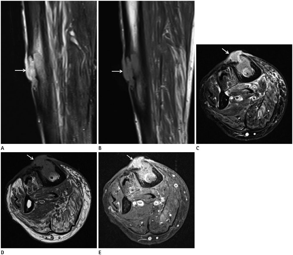
Fig. 3 MR images of the right lower leg. Sagittal T2-weighted fat suppressed image (A), sagittal T1-weighted image (B), axial T2-weighted fat suppressed image (C), axial T1-weighted image (D) and T1-weighted fat-suppressed image with gadolinium enhancement (E). Hyperintense soft tissue mass is seen on the anterior aspect of tibia on T2-weighted images (A, C, arrows). The lesion is isointense or slightly hyperintense than the muscle on T1-weighted image (B, D, arrows). Soft tissue mass extends through the cortical defect and then protrudes over subcutaneous tissue and skin (C, arrow). After enhancement, this lesion shows heterogeneously strong enhancement (E, arrow). Surrounding bone marrow edema is also noted. Edema and mild enhancement are also seen in the anterior and deep posterior compartment muscles of the lower leg, and on the anteromedial aspect of subcutaneous tissue and skin.
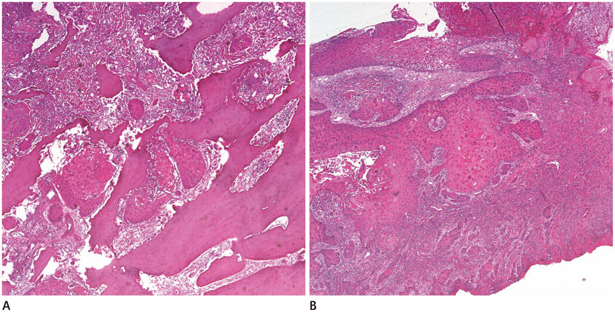
Fig. 4 Pathology. Squamous cell carcinoma is noted between bone fragments (A) and skin (B) (hematoxylin and eosin stain, × 40).
Reference
-
1. Panteli M, Puttaswamaiah R, Lowenberg DW, Giannoudis PV. Malignant transformation in chronic osteomyelitis: recognition and principles of management. J Am Acad Orthop Surg. 2014; 22:586–594.2. Treves N, Pack GT. The development of cancer in burn scars. Surg Gynecol Obstet. 1930; 51:749–782.3. Meaume S, Fromantin I, Teot L. Neoplastic wounds and degenerescence. J Tissue Viability. 2013; 22:122–130.4. Smith J, Mello LF, Nogueira Neto NC, Meohas W, Pinto LW, Campos VA, et al. Malignancy in chronic ulcers and scars of the leg (Marjolins' ulcer): a study of 21 patients. Skeletal Radiol. 2001; 30:331–337.5. Alami M, Mahfoud M, El Bardouni A, Berrada MS, El Yaacoubi M. Squamous cell carcinoma arising from chronic osteomyelitis. Acta Orthop Traumatol Turc. 2011; 45:144–148.6. Chiang KH, Chou AS, Hsu YH, Lee SK, Lee CC, Yen PS, et al. Marjolin's ulcer: MR appearance. AJR Am J Roentgenol. 2006; 186:819–820.7. Sawhney S, Jain R, Kakaria A, Chopra P. Marjolin's ulcer: radiographic and magnetic resonance appearances in two cases. Sultan Qaboos Univ Med J. 2009; 9:162–166.8. Luchs JS, Hines J, Katz DS, Athanasian EA. MR imaging of squamous cell carcinoma complicating chronic osteomyelitis of the femur. AJR Am J Roentgenol. 2002; 178:512–513.9. Ogawa B, Chen M, Margolis J, Schiller FJ, Schnall SB. Marjolin's ulcer arising at the elbow: a case report and literature review. Hand (N Y). 2006; 1:89–93.10. Shimose S, Sugita T, Kubo T, Matsuo T, Nobuto H, Ochi M. Differential diagnosis between osteomyelitis and bone tumors. Acta Radiol. 2008; 49:928–933.
- Full Text Links
-
- Actions
-
Cited
- CITED
-
- Close
- Share
-
- Similar articles
-
- Squamous Cell Carcinoma Arising from Chronic Osteomyelitis: A report of four Cases
- Squamous Cell Carcinoma Arising From Chronic Osteomyelitis: A Report of One Case
- Direct Bone Invasion of the Squamous Cell Carcinoma Arising from Chronic Osteomyelitis and Burn Scar: Report of 4 Cases
- A Case of Metastatic Cutaneous Squamous Cell Carcinoma Arising in Chronic Osteomyelitic Focus
- Squamous Cell Carcinoma Arising from Chronic Osteomyelitic Sinus: A Report of Three Cases
