Ann Dermatol.
2016 Jun;28(3):400-401. 10.5021/ad.2016.28.3.400.
Blue Toe Syndrome as an Early Sign of Disseminated Intravascular Coagulation
- Affiliations
-
- 1Department of Dermatology, VHS Medical Center, Seoul, Korea. choikohy@gmail.com
- KMID: 2164656
- DOI: http://doi.org/10.5021/ad.2016.28.3.400
Abstract
- No abstract available.
Figure
-

Fig. 1 Blue to purple discoloration with petechiae on the right foot.
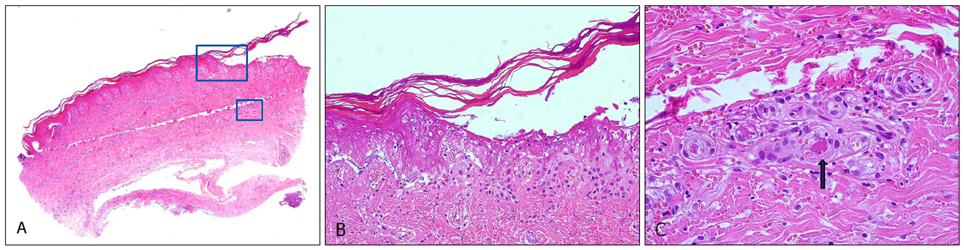
Fig. 2 (A) Scanning view (H&E, ×40). (B) Ischemic necrosis of epidermis, and red blood cell extravasation (H&E, ×200). (C) Eosinophilic fibrinoid thrombi in medium-sized vessels (arrow) and leukocytoslasis (H&E, ×400).
Reference
-
1. Chadachan V, Dean SM, Eberhardt RT. Cutaneous changes in peripheral arterial vascular disease. In : Goldsmith LA, Katz SI, Gilchrest BA, Paller AS, Leffell DJ, Wolff K, editors. Fitzpatrick's dermatology in general medicine. 8th ed. New York: McGraw Hill;2012. p. 2094–2110.2. Hirschmann JV, Raugi GJ. Blue (or purple) toe syndrome. J Am Acad Dermatol. 2009; 60:1–20.
Article3. Tschetter AJ, Liu V, Wanat KA. Cutaneous polyarteritis nodosa presenting as a solitary blue toe. J Am Acad Dermatol. 2014; 71:e95–e97.
Article4. Thornsberry LA, LoSicco KI, English JC 3rd. The skin and hypercoagulable states. J Am Acad Dermatol. 2013; 69:450–462.
Article5. Davis MP, Byrd J, Lior T, Rooke TW. Symmetrical peripheral gangrene due to disseminated intravascular coagulation. Arch Dermatol. 2001; 137:139–140.
- Full Text Links
-
- Actions
-
Cited
- CITED
-
- Close
- Share
-
- Similar articles
-
- Spontaneous Subdural Hematoma Associated with Disseminated Intravascular Coagulation in Patient with Cancer
- Disseminated intravascular coagulation
- Heparin Therapy for Disseminated Intravascular Coagulation in Childhood
- Occlusion o Left Middle Cerebral Artery Manifested as Disseminated Intravascular Coagulation
- A case of disseminated intravascular coagulation and acute renal insufficiency induced by falciparum malaria
