Computed tomographic bronchioarterial ratio for brachycephalic dogs without pulmonary disease
- Affiliations
-
- 1College of Veterinary Medicine and the Research Institute for Veterinary Science, Seoul National University, Seoul 151-742, Korea. heeyoon@snu.ac.kr
- 2Irion Animal Hospital, Seoul 135-100, Korea.
- 3College of Veterinary Medicine, Chonnam National University, Gwangju 500-757, Korea.
- KMID: 2164532
- DOI: http://doi.org/10.4142/jvs.2015.16.2.221
Abstract
- The bronchoarterial (BA) ratio measured with computed tomography is widely used in human medicine to diagnose bronchial dilation or collapse. Although use of the BA ratio in veterinary medicine has been recently studied, this has not been evaluated in brachycephalic dogs predisposed to bronchial diseases including bronchial collapse. The purpose of this study was to establish BA ratios for brachycephalic dogs and compare the values with those of non-brachycephalic dogs. Twenty-three brachycephalic dogs and 15 non-brachycephalic dogs without clinical pulmonary disease were evaluated. The BA ratio of the lobar bronchi in the left and right cranial as well as the right middle, left, and right caudal lung lobes was measured. No significant difference in mean BA ratio was observed between lung lobes or the individual animals (p = 0.148). The mean BA ratio was 1.08 +/- 0.10 (99% CI = 0.98~1.18) for brachycephalic dogs and 1.51 +/- 0.05 (99% CI = 1.46~1.56) for the non-brachycephalic group. There was a significant difference between the mean BA ratios of the brachycephalic and non-brachycephalic groups (p = 0.00). Defining the normal limit of the BA ratio for brachycephalic breeds may be helpful for diagnosing bronchial disease in brachycephalic dogs.
MeSH Terms
Figure
-
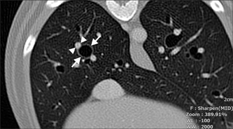
Fig. 1 Computed tomography (CT) image of the thorax of a brachycephalic dog at the level of the middle section of the ninth rib. The internal diameter of the right caudal bronchial lumen and diameter of the corresponding pulmonary artery in the cross-section were measured to determine the BA ratio. An arrow indicates the measured bronchus in the right caudal lung lobe while an arrowhead indicates the corresponding pulmonary artery.
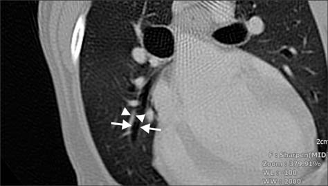
Fig. 2 CT image of the thorax of a dog at the level of the middle section of the seventh rib showing the right middle bronchus in the long-axis orientation. The internal diameter of the bronchial lumen and pulmonary artery diameter were measured to determine the BA ratio. An arrow indicates the measured diameter of the right middle lobar bronchus and an arrowhead indicates the corresponding pulmonary artery.

Fig. 3 Analysis of variation among the five lobes. No significant differences were detected among the BA ratios for the lobes (p = 0.148). LCR: left cranial lobe, LCA: left caudal lobe, RCR: right cranial lobe, RM: right middle lobe, RCA: right caudal lobe.
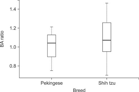
Fig. 4 Welch sample t-test results for the Shih-tzu and Pekingese dogs. There was no difference between the two breeds (p = 0.59).
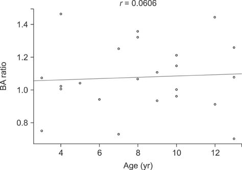
Fig. 5 Linear regression analysis of age and BA ratios. There is no correlation between BA ratio and age.
Reference
-
1. Adamama-Moraitou KK, Pardali D, Day MJ, Prassinos NN, Kritsepi-Konstantinou M, Patsikas MN, Rallis TS. Canine bronchomalacia: a clinicopathological study of 18 cases diagnosed by endoscopy. Vet J. 2012; 191:261–266.
Article2. Cannon MS, Wisner ER, Johnson LR, Kass PH. Computed tomography bronchial lumen to pulmonary artery diameter ratio in dogs without clinical pulmonary disease. Vet Radiol Ultrasound. 2009; 50:622–624.
Article3. De Lorenzi D, Bertoncello D, Drigo M. Bronchial abnormalities found in a consecutive series of 40 brachycephalic dogs. J Am Vet Med Assoc. 2009; 235:835–840.
Article4. Johnson LR, Pollard RE. Tracheal collapse and bronchomalacia in dogs: 58 cases (7/2001-1/2008). J Vet Intern Med. 2010; 24:298–305.
Article5. Kim SJ, Im JG, Kim IO, Cho ST, Cha SH, Park KS, Kim DY. Normal bronchial and pulmonary arterial diameters measured by thin section CT. J Comput Assist Tomogr. 1995; 19:365–369.
Article6. Matsuoka S, Uchiyama K, Shima H, Ueno N, Oish S, Nojiri Y. Bronchoarterial ratio and bronchial wall thickness on high-resolution CT in asymptomatic subjects: correlation with age and smoking. AJR Am J Roentgenol. 2003; 180:513–518.
Article7. Reid LE, Dillon AR, Hathcock JT, Brown LA, Tillson M, Wooldridge AA. High-resolution computed tomography bronchial lumen to pulmonary artery diameter ratio in anesthetized ventilated cats with normal lungs. Vet Radiol Ultrasound. 2012; 53:34–37.
Article
- Full Text Links
-
- Actions
-
Cited
- CITED
-
- Close
- Share
-
- Similar articles
-
- Computed tomographic features of gastric and esophageal content in dogs undergoing CT myelography and factors influencing the presence of esophageal fluid
- Virtual computed tomographic bronchoscopy: normal bronchial anatomy in six dogs
- Computed Tomographic Dacfyocystography using Rayvist(R)
- Sodium nitroprusside on acute cardiogenic pulmonary edema in dogs: case reports
- Computed tomographic anatomy of hepatic artery in normal beagle dogs
