Multiple Muscular Variations in the Neck, Upper Extremity, and Lower Extremity Biased toward the Left Side of a Single Cadaver
- Affiliations
-
- 1Brain Korea 21 PLUS Project for Medical Science, Yonsei University, Seoul, Korea.
- 2Department of Anatomy, Yonsei University College of Medicine, Seoul, Korea. leehy@yuhs.ac
- 3Department of Anatomy, Konkuk University College of Medicine, Seoul, Korea.
- 4Department of Anatomy, Gachon University School of Medicine, Incheon, Korea.
- 5Department of Anatomy, School of Medicine, Keimyung University, Daegu, Korea.
- KMID: 2164461
- DOI: http://doi.org/10.3346/jkms.2015.30.4.502
Abstract
- Although numerous reports have found accessory or supernumerary muscles throughout the human body, multiple appearances of these variations biased toward one side of body are rare. We report a 76-yr-old male cadaver with an accessory head of the biceps brachii and palmaris profundus, and a muscular slip between the biceps femoris and semitendinosus on the left side in addition to a bilateral accessory belly of the digastric muscle. No remarkable nervous, vascular, or visceral variation accompanied these variations. An interruption of normal somitogenesis or myogenesis may be a cause of these variations.
Keyword
Figure
-
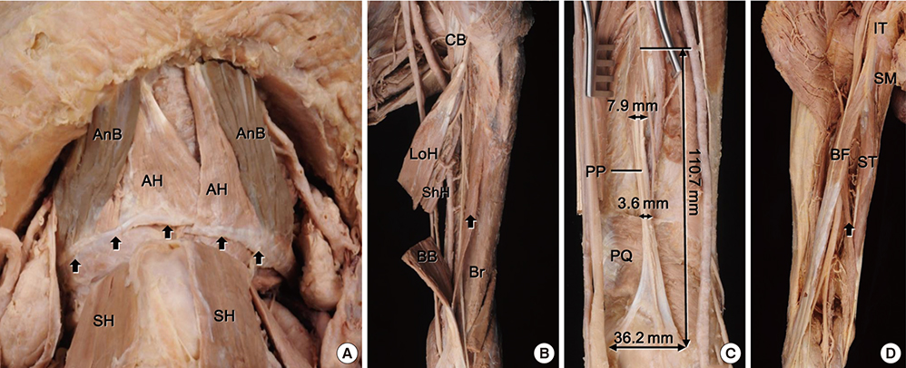
Fig. 1 Multiple muscular variations in a single cadaver. (A) accessory bellies (ABs) of the digastric muscles. These muscles were an anterior aspect of a fibrous band (arrows) between the intermediate tendons of digastric muscles on both sides. (B) third head of the biceps brachii muscle (arrow) in the left arm. This muscle originated at the humeral shaft inferior to the insertional site of the coracobrachialis muscle (CB). (C) palmaris profundus (PP) in the left forearm. The aberrant muscle originated from the interosseous membrane near the middle part of ulnar shaft and insert at the styloid processes of the radius and ulna. (D) aberrant muscular slip (arrow) between the biceps femoris and semitendinosus muscles (BF and ST) originated together with the long head of the BF about 127.7 mm lower than the ischial tuberosity (IT) and joined the ST at 110.5 mm above the insertion. AnB, anterior belly; Br, brachialis; BB, biceps brachii; LoH, long head of biceps brachii; PQ, pronator quadratus; SH, sternohyoid muscle; ShH, short head of biceps brachii; SM, semimembranosus.
Reference
-
1. De-Ary-Pires B, Ary-Pires R, Pires-Neto MA. The human digastric muscle: patterns and variations with clinical and surgical correlations. Ann Anat. 2003; 185:471–479.2. Fujimura A, Onodera M, Feng XY, Osawa T, Nara E, Nagato S, Matsumoto Y, Sasaki N, Nozaka Y. Abnormal anterior belly of the digastric muscle: a proposal for the classification of abnormalities. Anat Sci Int. 2003; 78:185–188.3. Sargon MF, Onderoğlu S, Sürücü HS, Bayramoğlu A, Demiryürek DD, Oztürk H. Anatomic study of complex anomalies of the digastric muscle and review of the literature. Okajimas Folia Anat Jpn. 1999; 75:305–313.4. Rodríguez-Niedenführ M, Vazquez T, Choi D, Parkin I, Sañudo JR. Supernumerary humeral heads of the biceps brachii muscle revisited. Clin Anat. 2003; 16:197–203.5. Reimann AF, Daseler EH, Anson BJ, Beaton LE. The palmaris longus muscle and tendon. A study of 1600 extremities. Anat Rec. 1944; 89:495–505.6. Seelaus HK. On certain muscle anomalies of the lower extremity. Anat Rec. 1927; 35:185–191.7. Sinav A, Gümüşalan Y, Arifoğlu Y, Onderoğlu S. Accessory muscular bundles arising from biceps femoris muscle. Kaibogaku Zasshi. 1995; 70:245–247.8. Mangalagiri AS, Razvi MR. Variations in the anterior belly of diagastric. Int J Health Sci (Qassim). 2009; 3:257–262.9. Yamazaki Y, Shibata M, Ushiki T, Isokawa K, Sato N. Bilateral, asymmetric anomalies of the anterior bellies of digastric muscles. J Oral Sci. 2011; 53:523–527.10. Rani A, Chopra J, Rani A, Verma RK. Duplicated anterior belly of the digastric muscle. Singapore Med J. 2013; 54:e131–e132.11. Abuel-Makarem SM, Ibrahim AF, Darwish HH. Absence of musculocutaneous nerve associated with a third head of biceps brachii muscle and entrapment of ulnar nerve. Neurosciences (Riyadh). 2007; 12:340–342.12. Yoshida Y, Yasutaka S, Seki Y. [Flexor radialis profundus and palmaris profundus muscles in man]. Kaibogaku Zasshi. 1983; 58:59–67.13. Pirola E, Hébert-Blouin MN, Amador N, Amrami KK, Spinner RJ. Palmaris profundus: one name, several subtypes, and a shared potential for nerve compression. Clin Anat. 2009; 22:643–648.14. Widmer CG, Morris-Wiman J. Limb, respiratory, and masticatory muscle compartmentalization: developmental and hormonal considerations. Prog Brain Res. 2010; 187:63–80.15. Kurose T, Asai Y, Mori E, Daitoku D, Kawamata S. Distribution and change of collagen types I and III and elastin in developing leg muscle in rat. Hiroshima J Med Sci. 2006; 55:85–91.16. Hootnick DR, Levinsohn EM, Crider RJ, Packard DS Jr. Congenital arterial malformations associated with clubfoot. A report of two cases. Clin Orthop Relat Res. 1982; 160–163.17. Sodre H, Bruschini S, Mestriner LA, Miranda F Jr, Levinsohn EM, Packard DS Jr, Crider RJ Jr, Schwartz R, Hootnick DR. Arterial abnormalities in talipes equinovarus as assessed by angiography and the Doppler technique. J Pediatr Orthop. 1990; 10:101–104.18. Schnorrer F, Dickson BJ. Muscle building; mechanisms of myotube guidance and attachment site selection. Dev Cell. 2004; 7:9–20.19. Brent AE. Somite formation: where left meets right. Curr Biol. 2005; 15:R468–R470.
- Full Text Links
-
- Actions
-
Cited
- CITED
-
- Close
- Share
-
- Similar articles
-
- Coexistence of Porokeratosis of Mibelli with Linear Porokeratosis
- Upper Extremity Deep Vein Thrombosis after Clavicle Fracture and Immobilization
- A Case of Cutis Marmorata Telangiectatica Congenits
- Motor Evoked Potentials of Upper and Lower Extremities by Magnetic Stimulation in Hemiparesis
- Two Cases of Partial Unilateral Lentiginosis
