Massive Upper Gastrointestinal Bleeding from Multiple Gastrointestinal Stromal Tumor in a Neurofibromatosis Patient
- Affiliations
-
- 1Division of Gastroenterology, Department of Internal Medicine, College of Medicine, The Catholic University of Korea, Seoul, Korea. jwchulkr@catholic.ac.kr
- KMID: 2164436
- DOI: http://doi.org/10.4166/kjg.2014.64.5.307
Abstract
- No abstract available.
MeSH Terms
-
Endoscopy, Digestive System
Gastrointestinal Hemorrhage/*etiology
Gastrointestinal Stromal Tumors/complications/*diagnosis/radionuclide imaging
Humans
Jejunum/pathology
Male
Middle Aged
Neurofibromatosis 1/complications/*diagnosis/pathology
Proto-Oncogene Proteins c-kit/metabolism
Tomography, X-Ray Computed
Proto-Oncogene Proteins c-kit
Figure
-
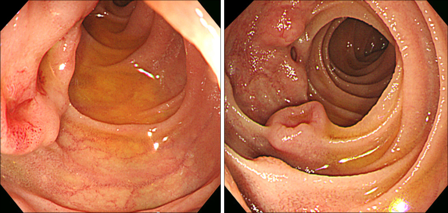
Fig. 1. Initial esophagogastroduod-enoscopy. Mass-like lesions with overlying normal mucosa and central ulceration are seen in the 2nd and 3rd portion of the duodenum.

Fig. 2. Abdominal computed tomography. Two well-enhancing lesions (arrows) are observed in the third portion of the duodenum.
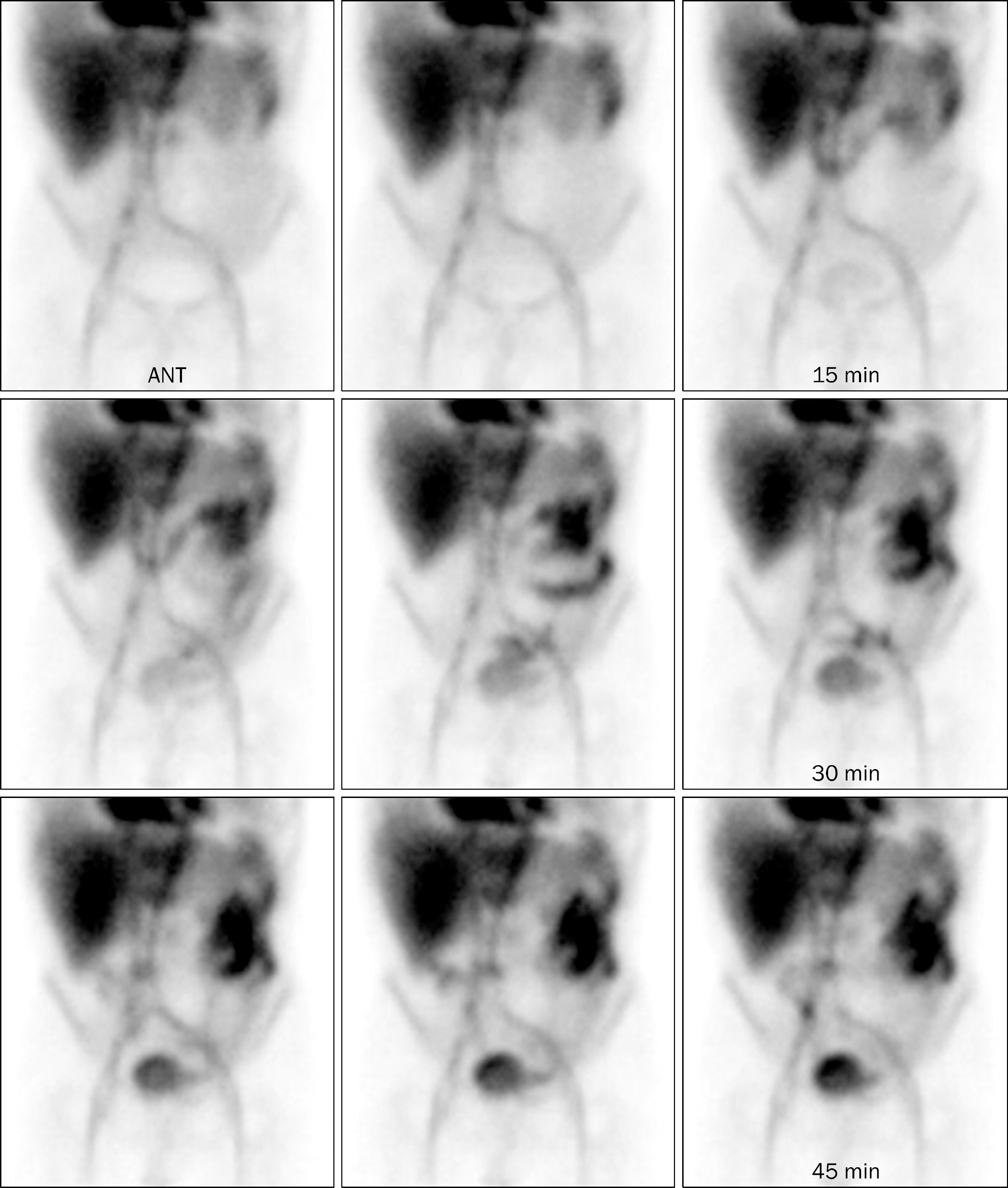
Fig. 3. Gastrointestinal bleeding scan with Tc-99m labeled red blood cell (RBC). An area of accumulation of Tc-99m RBC is noted at left upper abdomen with increased amount of activity and distal migration, suggesting bleeding from upper jejunum. ANT, anterior.

Fig. 4. Histologic examination of the biopsy specimen. (A) Spindle shaped cells arranged in fascicular pattern are seen (H&E, ×100). (B) Tumor cells are positive for CD 117 on immunohistochemical staining (×100).
Reference
-
References
1. Riccardi VM. Neurofibromatosis: clinical heterogeneity. Curr Probl Cancer. 1982; 7:1–34.
Article2. Relles D, Baek J, Witkiewicz A, Yeo CJ. Periampullary and duodenal neoplasms in neurofibromatosis type 1: two cases and an updated 20-year review of the literature yielding 76 cases. J Gastrointest Surg. 2010; 14:1052–1061.
Article3. North K. Neurofibromatosis type 1. Am J Med Genet. 2000; 97:119–127.
Article4. Basile U, Cavallaro G, Polistena A, et al. Gastrointestinal and retroperitoneal manifestations of type 1 neurofibromatosis. J Gastrointest Surg. 2010; 14:186–194.
Article5. Kim ET, Namgung H, Shin HD, et al. Oncologic manifestations of neurofibromatosis type 1 in Korea. J Korean Surg Soc. 2012; 82:205–210.
Article6. Gottfried ON, Viskochil DH, Couldwell WT. Neurofibromatosis Type 1 and tumorigenesis: molecular mechanisms and therapeutic implications. Neurosurg Focus. 2010; 28:E8.
Article7. Andersson J, Sihto H, Meis-Kindblom JM, Joensuu H, Nupponen N, Kindblom LG. NF1-associated gastrointestinal stromal tumors have unique clinical, phenotypic, and genotypic characteristics. Am J Surg Pathol. 2005; 29:1170–1176.
Article8. Miettinen M, Lasota J. Gastrointestinal stromal tumors-defi-nition, clinical, histological, immunohistochemical, and molecular genetic features and differential diagnosis. Virchows Arch. 2001; 438:1–12.9. Miettinen M, Fetsch JF, Sobin LH, Lasota J. Gastrointestinal stromal tumors in patients with neurofibromatosis 1: a clinicopathologic and molecular genetic study of 45 cases. Am J Surg Pathol. 2006; 30:90–96.10. Choi W, Hong SD, Kim HN, et al. Two cases of neuroendocrine carcinoma and GIST in a patient with neurofibromatosis type 1. Korean J Med. 2011; 81:786–791.11. Seo SO, Oh HJ, Kim KH, et al. A case of duodenal GIST accompanied with neurofibromatosis-1, presenting with gastrointestinal bleeding. Korean J Gastrointest Endosc. 2007; 35:254–257.12. Han SH, Park SH, Cho GH, et al. Malignant gastrointestinal stromal tumor in a patient with neurofibromatosis type 1. Korean J Intern Med. 2007; 22:21–23.
Article
- Full Text Links
-
- Actions
-
Cited
- CITED
-
- Close
- Share
-
- Similar articles
-
- A Case of Massive Bleeding from Jejunal Stromal Tumor Diagnosed by Intraoperative Enteroscopy: A Case of Jejunal Stromal Tumor Bleeding
- Solitary Malignant Gastrointestinal Stromal Tumor Associated with a Neurofibromatosis Type I
- A Case of Duodenal GIST Accompanied with Neurofibromatosis-1, Presenting with Gastrointestinal Bleeding
- A Case of Chronic Recurrent Small Bowel Bleeding in Neurofibromatosis Type 1 Diagnosed by Capsule Endoscopy
- A Case Report of a Bleeding Duodenal Gastro-Intestinal Stromal Tumor and its Emergent Management
