Korean J Gastroenterol.
2014 Nov;64(5):298-301. 10.4166/kjg.2014.64.5.298.
A Case of Fascioliasis in the Intrahepatic Duct with Concurrent Clonochiasis
- Affiliations
-
- 1Department of Internal Medicine, Dongkang Medical Center, Ulsan, Korea. sky8170@lycos.co.kr
- KMID: 2164434
- DOI: http://doi.org/10.4166/kjg.2014.64.5.298
Abstract
- The main causes of biliary obstruction are stones and cancers. Fascioliasis is a very rare case which causes biliary obstruction. Fascioliasis is a zoonosis caused by Fasciola hepatica which infects herbivores like sheep and cattle. F. hepatica lives in the biliary system or the liver parenchyma of a host. In Korea, the occurrence of this infection in human is very rare and only few cases have been reported. A 32-year-old male presented with upper abdominal pain and jaundice. His laboratory finding revealed elevated liver transaminases. Abdomen CT scan showed mild left intrahepatic bile duct dilatation. On ERCP, adult F. hepatica worms were found and were thus removed. Concurrently, clonorchiasis was diagnosed by stool exam and serologic enzyme-linked immunosorbent assay test. Clonorchiasis was treated with praziquantel. Herein, we report a case of intrahepatic bile duct dilatation due to F. hepatica infection with concurrent Clonorchis sinensis infestation.
Keyword
MeSH Terms
-
Adult
Animals
Anthelmintics/therapeutic use
Benzimidazoles/therapeutic use
Bile Ducts, Intrahepatic
Cholangiopancreatography, Endoscopic Retrograde
Clonorchiasis/complications/*diagnosis/drug therapy
Clonorchis sinensis/immunology/isolation & purification
Enzyme-Linked Immunosorbent Assay
Fasciola/isolation & purification
Fascioliasis/complications/*diagnosis/parasitology
Humans
Liver/enzymology
Male
Praziquantel/therapeutic use
Tomography, X-Ray Computed
Transaminases/metabolism
Anthelmintics
Benzimidazoles
Praziquantel
Transaminases
Figure
-
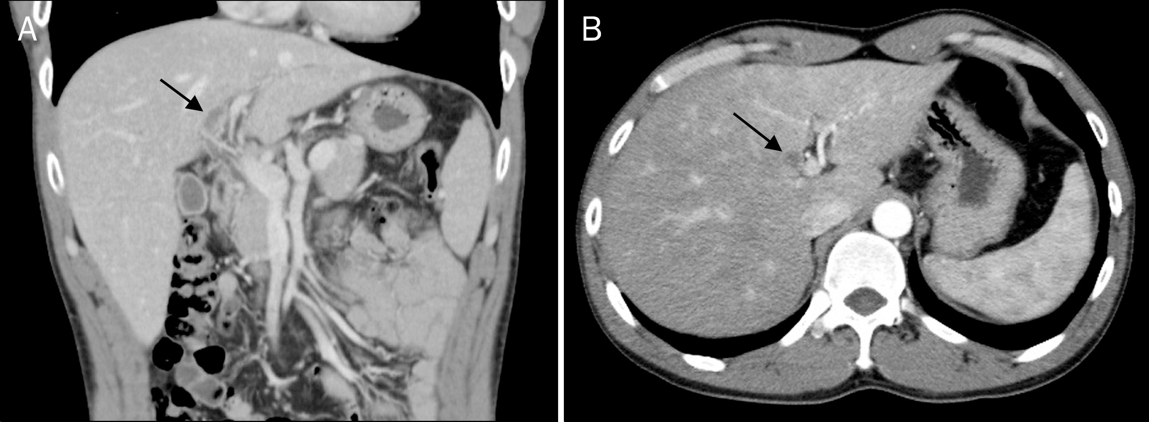
Fig. 1. Abdomen computed tomography scan findings. On (A) coronal reconstructed image and (B) axial scan, mild left intrahepatic bile duct dilatation is noted but no definite obstructive lesion is observed (arrows).
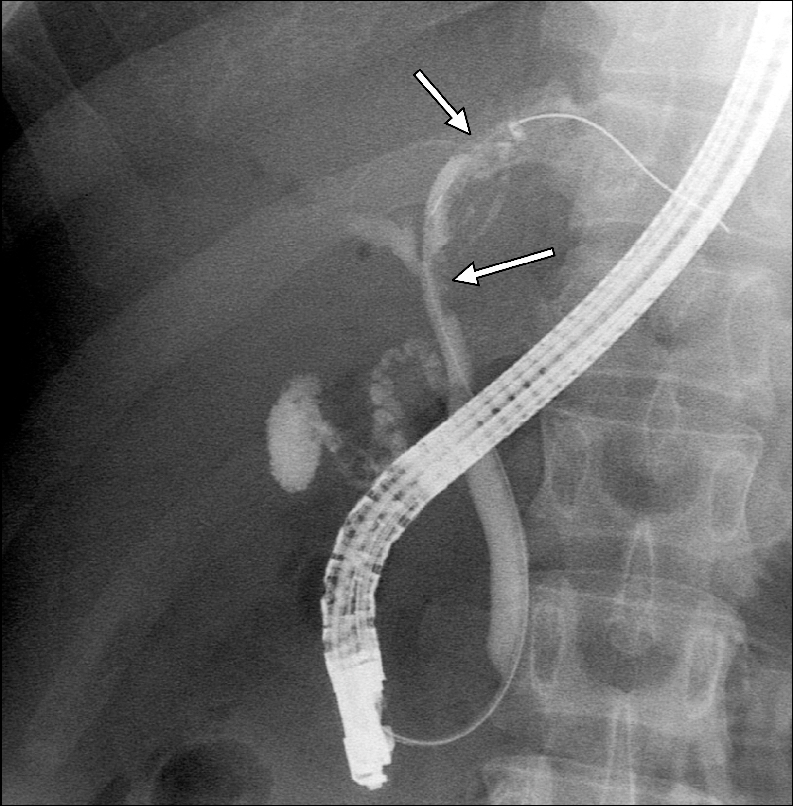
Fig. 2. Endoscopic retrograde cholangiopancreatography shows multiple filling defects (arrows) in the left intrahepatic bile duct.
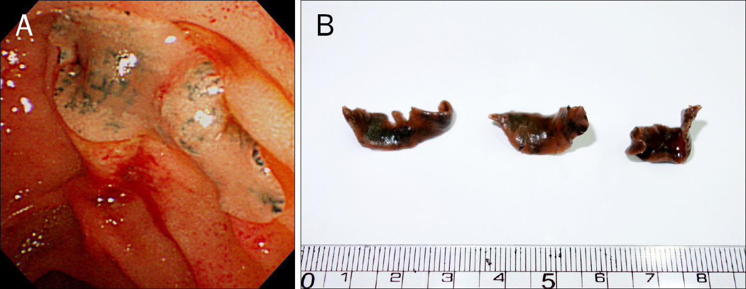
Fig. 3. (A) The adult Fasciola hepatica worms were removed by endoscopic balloon catheter sweeping from the left intrahepatic bile duct. (B) Three adult F. hepatica worms measuring abount 25–30 mm in length are shown.
Reference
-
References
1. Cho SY, Seo BS, Kim YI, Won CK, Cho SK. A case of human fascioliasis in Korea. Korean J Parasitol. 1976; 14:147–152.
Article2. Paik JW, Choi CS, Choi YK. A case of fascioliasis in the common bile duct. Korean J Gastroenterol. 1997; 30:553–558.3. Kim CK, Lee HY, Paik SW, et al. A case of fascioliasis presented as a single, large hepatic mass. Korean J Gastroenterol. 1997; 30:689–694.4. Woo SY, Jung HJ, Kim WT, et al. A case of acute pancreatitis associated with fasciola hepatica. Korean J Gastrointest Endosc. 2006; 33:183–186.5. Marcos LA, Terashima A, Gotuzzo E. Update on hepatobiliary flukes: fascioliasis, opisthorchiasis and clonorchiasis. Curr Opin Infect Dis. 2008; 21:523–530.
Article6. Rowan SE, Levi ME, Youngwerth JM, Brauer B, Everson GT, Johnson SC. The variable presentations and broadening geo-graphic distribution of hepatic fascioliasis. Clin Gastroenterol Hepatol. 2012; 10:598–602.
Article7. Shon JH, Cho KB, Hwang JS, et al. A case of liver abscess associated with fascioliasis diagnosed by MRI. Korean J Hepatol. 2001; 7:90–94.8. Choi JH, Kim DS, Choi WH, Lee TS, Chung DI, Choi DW. A human case of hepatic fascioliasis accompanied by egg granulomas in common bile duct lymph node. Korean J Pathol. 1991; 25:250–255.9. Dusak A, Onur MR, Cicek M, Firat U, Ren T, Dogra VS. Radiologi-cal imaging features of fasciola hepatica infection: a pictorial review. J Clin Imaging Sci. 2012; 2:2.10. Chang R, Kim BH, Kim HJ, et al. Biliary tract & pancreas: a case of fascioliasis diagnosed by ERCP. Korean J Gastrointest Endosc. 1997; 17:105–109.11. Ha SW, Chang HH, Kim SW, et al. A case of fascioliasis diagnosed by endoscopic nasobiliary drainage fluid examination. Korean J Med. 2007; 72:658–662.12. Chai JY. Praziquantel treatment in trematode and cestode infections: an update. Infect Chemother. 2013; 45:32–43.
Article13. Keiser J, Engels D, Büscher G, Utzinger J. Triclabendazole for the treatment of fascioliasis and paragonimiasis. Expert Opin Investig Drugs. 2005; 14:1513–1526.
Article14. Nagano I, Pei F, Wu Z, et al. Molecular expression of a cysteine proteinase of Clonorchis sinensis and its application to an en-zyme-linked immunosorbent assay for immunodiagnosis of clonorchiasis. Clin Diagn Lab Immunol. 2004; 11:411–416.15. Zhao QP, Moon SU, Lee HW, et al. Evaluation of Clonorchis sinensis recombinant 7-kilodalton antigen for serodiagnosis of clonorchiasis. Clin Diagn Lab Immunol. 2004; 11:814–817.
