Characteristics of the molar surface after removal of cervical enamel projections: comparison of three different rotating instruments
- Affiliations
-
- 1Department of Periodontology, Wonkwang University Daejeon Dental Hospital, Wonkwang University School of Dentistry, Daejeon, Korea. seongnyum@wonkwang.ac.kr
- KMID: 2161987
- DOI: http://doi.org/10.5051/jpis.2016.46.2.107
Abstract
- PURPOSE
The aim of this study was to evaluate and compare tooth surface characteristics in extracted human molars after cervical enamel projections (CEPs) were removed with the use of three rotating instruments.
METHODS
We classified 60 extracted molars due to periodontal lesion with CEPs into grade I, II, or III, according to the Masters and Hoskins' criteria. Each group contained 20 specimens. Three rotating instruments were used to remove the CEPs: a piezoelectric ultrasonic scaler, a periodontal bur, and a diamond bur. Tooth surface characteristics before and after removal of the projections were then evaluated with scanning electron microscopy (SEM). We analyzed the characteristics of the tooth surfaces with respect to roughness and whether the enamel projections had been completely removed.
RESULTS
In SEM images, surfaces treated with the diamond bur were smoothest, but this instrument caused considerable harm to tooth structures near the CEPs. The piezoelectric ultrasonic scaler group produced the roughest surface but caused less harm to the tooth structure near the furcation. In general, the surfaces treated with the periodontal bur were smoother than those treated with the ultrasonic scaler, and the periodontal bur did not invade adjacent tooth structures.
CONCLUSIONS
For removal of grade II CEPs, the most effective instrument was the diamond bur. However, in removing grade III projections, the diamond bur can destroy both adjacent tooth structures and the periodontal apparatus. In such cases, careful use of the periodontal bur may be an appropriate substitute.
MeSH Terms
Figure
-
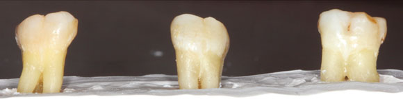
Figure 1 Extracted molars embedded in a plaster model.
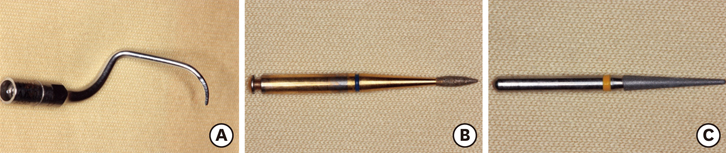
Figure 2 Rotary instruments used in this study: piezoelectric ultrasonic scaler tip (A), periodontal bur (B), and diamond bur (C).

Figure 3 Preparation of specimens 5×6×2 mm3 in size containing the cementoenamel junction.
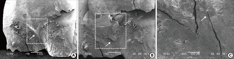
Figure 4 Representative SEM photographs of tooth surface before (A) and after (B and C) removal of grade II cervical enamel projections with the use of an ultrasonic scaler (A, B: 20×, C: 200×).

Figure 6 Representative SEM photographs of tooth surface before (A) and after (B and C) removal of grade I cervical enamel projections with the use of a periodontal bur (A, B: 20×, C: 200×).
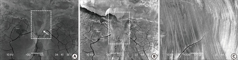
Figure 8 Representative SEM photographs of tooth surface before (A) and after (B and C) removal of grade II cervical enamel projections with the use of a diamond bur (A, B: 20×, C: 200×).
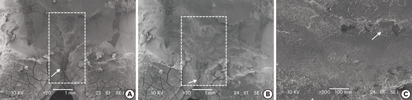
Figure 5 Representative SEM photographs of tooth surface before (A) and after (B and C) removal of grade III cervical enamel projections with the use of an ultrasonic scaler (A, B: 20×, C: 200×).
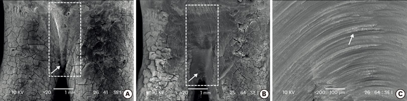
Figure 7 Representative SEM photographs of tooth surface before (A) and after (B and C) removal of grade III cervical enamel projections with the use of a periodontal bur (A, B: 20×, C: 200×).
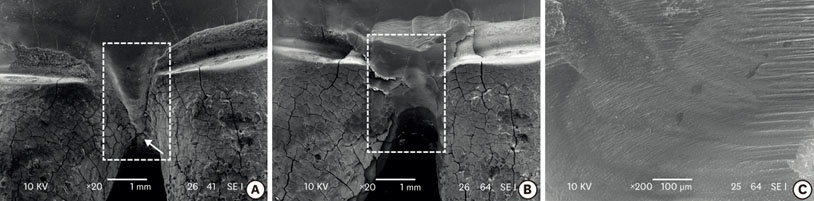
Figure 9 Representative SEM photographs of tooth surface before (A) and after (B and C) removal of grade III cervical enamel projections with the use of a diamond bur (A, B: 20×, C: 200×).
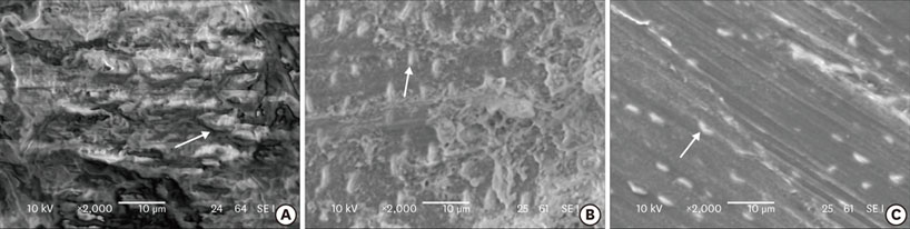
Figure 10 Representative SEM photographs of dentinal tubules. (A) Ultrasonic scaler group (2,000×). (B) Periodontal bur group (2,000×). (C) Diamond bur group (2,000×). Each arrowhead indicates the occluded entrance of dentinal tubules.
Reference
-
1. Nishihara T, Koseki T. Microbial etiology of periodontitis. Periodontol 2000. 2004; 36:14–26.
Article2. Attar NB, Phadnaik MB. Bilateral cervicoenamel projection and its management: A case report with lingual involvement. J Indian Soc Periodontol. 2009; 13:168–171.
Article3. Askenas BG, Fry HR, Davis JW. Cervical enamel projection with gingival fenestration in a maxillary central incisor: report of a case. Quintessence Int. 1992; 23:103–107.4. Masters DH, Hoskins SW Jr. Projection of cervical enamel into molar furcations. J Periodontol. 1964; 35:49–53.
Article5. Swan RH, Hurt WC. Cervical enamel projections as an etiologic factor in furcation involvement. J Am Dent Assoc. 1976; 93:342–345.
Article6. Atkinson SR. Changing dynamics of the growing face. Am J Orthod. 1949; 35:815–836.
Article7. Grewe JM, Meskin LH, Miller T. Cervical enamel projections: prevalence, location, and extent; with associated periodontal implications. J Periodontol. 1965; 36:460–465.
Article8. Blanchard SB, Derderian GM, Averitt TR, John V, Newell DH. Cervical enamel projections and associated pouch-like opening in mandibular furcations. J Periodontol. 2012; 83:198–203.
Article9. Carranza FA Jr, Jolkovsky DL. Current status of periodontal therapy for furcation involvements. Dent Clin North Am. 1991; 35:555–570.10. Hou GL, Tsai CC. Relationship between periodontal furcation involvement and molar cervical enamel projections. J Periodontol. 1987; 58:715–721.
Article11. Hou GL, Tsai CC. Cervical enamel projection and intermediate bifurcational ridge correlated with molar furcation involvements. J Periodontol. 1997; 68:687–693.
Article12. Machtei EE, Wasenstein SM, Peretz B, Laufer D. The relationship between cervical enamel projection and class II furcation defects in humans. Quintessence Int. 1997; 28:315–320.13. Bower RC. Furcation morphology relative to periodontal treatment. Furcation root surface anatomy. J Periodontol. 1979; 50:366–374.
Article14. Shiloah J, Kopczyk RA. Developmental variations of tooth morphology and periodontal disease. J Am Dent Assoc. 1979; 99:627–630.
Article15. Cho KY, Choi SM. Prevalence of cervical enamel projections and its relation to furcation involvement. J Korean Acad Periodontol. 1986; 16:96–97.16. Moskow BS. Some observations on radicular enamel. J Periodontol. 1971; 42:92–96.
Article17. Bissada NF, Abdelmalek RG. Incidence of cervical enamel projections and its relationship to furcation involvement in Egyptian skulls. J Periodontol. 1973; 44:583–585.
Article18. Bye FL, Ghilzan RS, Coffesse RG. Root surface roughness after the use of different modes of instrumentation. Int J Periodontics Restorative Dent. 1986; 6:36–47.19. Rosenberg RM, Ash MM Jr. The effect of root roughness on plaque accumulation and gingival inflammation. J Periodontol. 1974; 45:146–150.
Article20. Khatiblou FA, Ghodssi A. Root surface smoothness or roughness in periodontal treatment. A clinical study. J Periodontol. 1983; 54:365–367.
Article21. Stende GW, Schaffer EM. A comparison of ultrasonic and hand scaling. J Periodontol. 1961; 32:312–314.
Article22. Kawashima H, Sato S, Kishida M, Ito K. A comparison of root surface instrumentation using two piezoelectric ultrasonic scalers and a hand scaler in vivo. J Periodontal Res. 2007; 42:90–95.
Article23. Kishida M, Sato S, Ito K. Comparison of the effects of various periodontal rotary instruments on surface characteristics of root surface. J Oral Sci. 2004; 46:1–8.
Article24. Heo SR, Kim SA, Seo SR, Kim HS. A study on the loss of tooth substance and surface changes following root planing. J Korean Acad Periodontol. 1998; 28:351–369.
Article25. Eick S, Bender P, Flury S, Lussi A, Sculean A. In vitro evaluation of surface roughness, adhesion of periodontal ligament fibroblasts, and Streptococcus gordonii following root instrumentation with Gracey curettes and subsequent polishing with diamond-coated curettes. Clin Oral Investig. 2013; 17:397–404.
Article26. Aspriello SD, Piemontese M, Levrini L, Sauro S. Ultramorphology of the root surface subsequent to hand-ultrasonic simultaneous instrumentation during non-surgical periodontal treatments: an in vitro study. J Appl Oral Sci. 2011; 19:74–81.
Article27. Bower RC. Furcation morphology relative to periodontal treatment. Furcation entrance architecture. J Periodontol. 1979; 50:23–27.
Article
- Full Text Links
-
- Actions
-
Cited
- CITED
-
- Close
- Share
-
- Similar articles
-
- Effects of various clean-up techniques on enamel surface roughness
- 3D quantitative analysis and SEM qualitative analysis of natural antagonist enamel opposing CAD-CAM monolithic zirconia or lithium disilicate tooth-supported crowns versus enamel opposing natural enamel
- A comparative study of roughness of enamel surface to various interdental enamel stripping methods in vitro
- Relationship between mesiodistal width and enamel thickness in mandibular incisors
- A study of enamel demineralization related to bonded orthodontic bracket and improved method of enamel demineralization: in vivo study
