Multiple osteoblastomas in a child with Cushing syndrome due to bilateral adrenal micronodular hyperplasias
- Affiliations
-
- 1Department of Pediatrics, Seoul National University Children's Hospital, Seoul, Korea.
- 2Department of Pediatrics, Seoul National University Bundang Hospital, Seongnam, Korea. chyerim@hanmail.net
- 3Department of Pathology, Seoul National University Bundang Hospital, Seongnam, Korea.
- 4Department of Orthopaedic Surgery, Seoul National University Bundang Hospital, Seongnam, Korea.
- KMID: 2161307
- DOI: http://doi.org/10.6065/apem.2016.21.1.47
Abstract
- Adrenocorticotropin-independent adrenal hyperplasias are rare diseases, which are classified into macronodular (>1 cm) and micronodular (≤1 cm) hyperplasia. Micronodular adrenal hyperplasia is subdivided into primary pigmented adrenocortical disease and a limited or nonpigmented form 'micronodular adrenocortical disease (MAD)', although considerable morphological and genetic overlap is observed between the 2 groups. We present an unusual case of a 44-month-old girl who was diagnosed with Cushing syndrome due to MAD. She had presented with spotty pigmentation on her oral mucosa, lips and conjunctivae and was diagnosed with multiple bone tumors in her femur, pelvis and skull base at the age of 8 years. Her bone tumor biopsies were compatible with osteoblastoma. This case highlights the importance of verifying the clinicopathologic correlation in Cushing syndrome and careful follow-up and screening for associated diseases.
MeSH Terms
Figure
-

Fig. 1 Computed tomography of the adrenal gland. Horizontal view of the abdomen revealed 5-mm nodular thickening of the left adrenal gland and a nearly normal right adrenal gland (black arrow).

Fig. 2 Pathology of both adrenal glands. (A) The right adrenal gland measured 4 cm×3 cm×0.7 cm, and the left adrenal gland measured 5.5 cm×3.2 cm×1.5 cm. The adrenal glands are enlarged and have multiple micronodules on the cut surface (white arrows). Microscopically, circumscribed small nodules are observed in the adrenal cortex, and the nodule diameter usually does not exceed 0.5 cm. No pigment granules are observed. H&E staining (B: ×40; C: ×100).
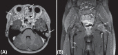
Fig. 3 Magnetic resonance imaging of brain and hip. (A) Horizontal view of the brain demonstrates multiple multilocular cystic bone lesions with internal hemorrhage and prominent wall enhancements involving the clivus, bifrontal skull base, bilateral sphenoid and left temporal bones (white arrows). (B) Coronal view of the hip with section at the femur head level revealed overall polyostotic bone tumors involving bilateral femurs as well as the ilium and sacrum (white arrows)

Fig. 4 Pathologic finding of osteoblastoma (H&E, ×200). The lesion consists of multinucleated giant cell, immature bone and osteoid deposition in fibrovascular stroma.
Reference
-
1. Magiakou MA, Mastorakos G, Oldfield EH, Gomez MT, Doppman JL, Cutler GB Jr, et al. Cushings syndrome in children and adolescents. Presentation, diagnosis, and therapy. N Engl J Med. 1994; 331:629–623. PMID: 8052272.
Article2. Nieman LK, Biller BM, Findling JW, Newell-Price J, Savage MO, Stewart PM, et al. The diagnosis of Cushing's syndrome: an Endocrine Society Clinical Practice Guideline. J Clin Endocrinol Metab. 2008; 93:1526–1540. PMID: 18334580.3. Sharma ST, Nieman LK, Feelders RA. Cushing's syndrome: epidemiology and developments in disease management. Clin Epidemiol. 2015; 7:281–293. PMID: 25945066.4. Boscaro M, Barzon L, Fallo F, Sonino N. Cushing's syndrome. Lancet. 2001; 357:783–791. PMID: 11253984.5. Stratakis CA, Boikos SA. Genetics of adrenal tumors associated with Cushing's syndrome: a new classification for bilateral adrenocortical hyperplasias. Nat Clin Pract Endocrinol Metab. 2007; 3:748–757. PMID: 17955016.6. Duan K, Gomez Hernandez K, Mete O. Clinicopathological correlates of adrenal Cushing's syndrome. J Clin Pathol. 2015; 68:175–186. PMID: 25425660.7. Batista DL, Riar J, Keil M, Stratakis CA. Diagnostic tests for children who are referred for the investigation of Cushing syndrome. Pediatrics. 2007; 120:e575–e586. PMID: 17698579.8. Carney JA, Gaillard RC, Bertherat J, Stratakis CA. Familial micronodular adrenocortical disease, Cushing syndrome, and mutations of the gene encoding phosphodiesterase 11A4 (PDE11A). Am J Surg Pathol. 2010; 34:547–555. PMID: 20351491.
Article9. Almeida MQ, Stratakis CA. Carney complex and other conditions associated with micronodular adrenal hyperplasias. Best Pract Res Clin Endocrinol Metab. 2010; 24:907–914. PMID: 21115159.
Article10. Carney JA, Boccon-Gibod L, Jarka DE, Tanaka Y, Swee RG, Unni KK, et al. Osteochondromyxoma of bone: a congenital tumor associated with lentigines and other unusual disorders. Am J Surg Pathol. 2001; 25:164–176. PMID: 11176065.11. Courcoutsakis NA, Tatsi C, Patronas NJ, Lee CC, Prassopoulos PK, Stratakis CA. The complex of myxomas, spotty skin pigmentation and endocrine overactivity (Carney complex): imaging findings with clinical and pathological correlation. Insights Imaging. 2013; 4:119–133. PMID: 23315333.
Article
- Full Text Links
-
- Actions
-
Cited
- CITED
-
- Close
- Share
-
- Similar articles
-
- A Case of Bilateral ACTH-independent Adrenal Adenomas with Cushing's Syndrome Treated by Ipsilateral Total and Contralateral Partial Laparoscopic Adrenalectomy
- Three Cases of Functioning Adrenocortical Tumor
- Cushing Syndrome Caused by ACTH-independent Macronodular Adrenal Hyperplasia
- A Case of Cushing's Syndrome in Pregnancy Secondary to an Adrenal Cortical Adenoma
- A Case of Black Adrenocortical Adenoma Causing Cushing's Syndrome with Contralateral Nonfuncioning Adenoma
