Robot-assisted laparoscopic retroperitoneal lymph node dissection for stage IIIb mixed germ cell testicular cancer after chemotherapy
- Affiliations
-
- 1Department of Urology, Kyung Hee University School of Medicine, Seoul, Korea. juro@khu.ac.kr
- KMID: 2160652
- DOI: http://doi.org/10.4111/kju.2015.56.7.540
Abstract
- Laparoscopic retroperitoneal lymph node dissection, especially when performed with the da Vinci Surgical System (Intuitive Surgical), has shown excellent cosmetic results with similar oncologic outcomes to those of open surgery. In this study, we present a case of robot-assisted retroperitoneal lymph node dissection performed in an 18-year-old man who was diagnosed with a stage IIIb mixed germ cell tumor and who was initially treated with radical orchiectomy, followed by chemotherapy. This case shows that robot-assisted retroperitoneal lymph node dissection is technically feasible, safe, and cosmetically favorable, even when performed on patients with high-stage disease or after chemotherapy.
MeSH Terms
-
Adolescent
Chemotherapy, Adjuvant
Humans
Laparoscopy/methods
Lymph Node Excision/*methods
Lymphatic Metastasis
Male
Neoplasm Staging
Neoplasms, Germ Cell and Embryonal/pathology/radiography/*secondary/therapy
Orchiectomy
Robotic Surgical Procedures/*methods
Testicular Neoplasms/pathology/radiography/*secondary/therapy
Tomography, X-Ray Computed
Figure
-
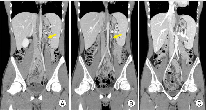
Fig. 1 Computed tomography images. (A) Before chemotherapy: a para-aortic lymph node is indicated by the yellow arrow. (B) After chemotherapy: the size of the lymph node was decreased after three cycles of bleomycin-etoposide-cisplatin chemotherapy; however, a remnant mass (yellow arrow) was detected. (C) At 18 months after retroperitoneal lymph node dissection, no lymph nodes were detected during follow-up.
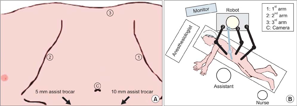
Fig. 2 (A) Port placement (C, camera port; 1, first robotic arm; 2, second robotic arm; 3, third robotic arm). (B) Robot dock, assistant, and nurse positioning.
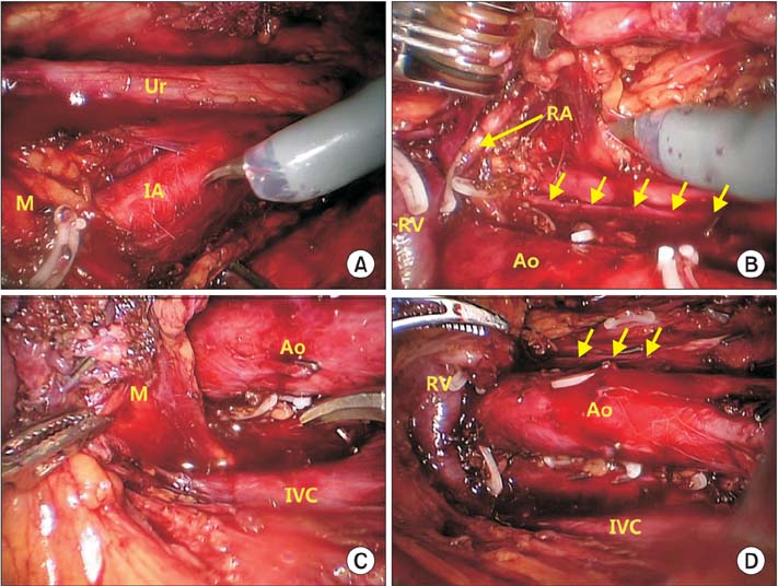
Fig. 3 Images from robot-assisted laparoscopic retroperitoneal lymph node dissection. First, the retroperitoneal space was exposed. (A) Lymph nodes were dissected from the level of the common iliac artery. (B) Dissection was continued to the hilar area. All of the paraaortic lymph nodes were removed. (C) Interaortocaval lymph nodes, which were not found on the previous computed tomography, were detected and removed. (D) After the removal of whole lymph nodes, skeletonized great vessels were detected. Yellow arrows on panels B and D indicate the sympathetic nerve chain along the lateral border of the aorta. Ur, ureter; M, mass; IA, common iliac artery; RA, renal artery; RV, renal vein; Ao, aorta; IVC, inferior vena cava. Scan this QR code to see the accompanying video, or visit www.kjurology.org or http://youtu.be/CLiPJHAQyCc.
Reference
-
1. Rassweiler JJ, Scheitlin W, Heidenreich A, Laguna MP, Janetschek G. Laparoscopic retroperitoneal lymph node dissection: does it still have a role in the management of clinical stage I nonseminomatous testis cancer? A European perspective. Eur Urol. 2008; 54:1004–1015.2. Davol P, Sumfest J, Rukstalis D. Robotic-assisted laparoscopic retroperitoneal lymph node dissection. Urology. 2006; 67:199.3. Williams SB, Lau CS, Josephson DY. Initial series of robot-assisted laparoscopic retroperitoneal lymph node dissection for clinical stage I nonseminomatous germ cell testicular cancer. Eur Urol. 2011; 60:1299–1302.4. Mir MC, Autorino R, Samarasekera D, Klink J, Stephenson AJ, Kaouk JH. Robot-assisted laparoscopic retroperitoneal lymph node dissection for left clinical stage I non-seminomatous germ cell testicular cancer: focus on port placement and surgical technique. Int J Urol. 2014; 21:212–214.5. Cheney SM, Andrews PE, Leibovich BC, Castle EP. Robot-assisted retroperitoneal lymph node dissection: technique and initial case series of 18 patients. BJU Int. 2015; 115:114–120.6. Shin YS, Kim HJ. Current management of testicular cancer. Korean J Urol. 2013; 54:2–10.7. Steiner H, Leonhartsberger N, Stoehr B, Peschel R, Pichler R. Postchemotherapy laparoscopic retroperitoneal lymph node dissection for low-volume, stage II, nonseminomatous germ cell tumor: first 100 patients. Eur Urol. 2013; 63:1013–1017.8. Palese MA, Su LM, Kavoussi LR. Laparoscopic retroperitoneal lymph node dissection after chemotherapy. Urology. 2002; 60:130–134.9. Calestroupat JP, Sanchez-Salas R, Cathelineau X, Rozet F, Galiano M, Smyth G, et al. Postchemotherapy laparoscopic retroperitoneal lymph node dissection in nonseminomatous germ-cell tumor. J Endourol. 2009; 23:645–650.10. Cost NG, DaJusta DG, Granberg CF, Cooksey RM, Laborde CE, Wickiser JE, et al. Robot-assisted laparoscopic retroperitoneal lymph node dissection in an adolescent population. J Endourol. 2012; 26:635–640.
- Full Text Links
-
- Actions
-
Cited
- CITED
-
- Close
- Share
-
- Similar articles
-
- Clinicopathologic and Oncological Outcomes in Korean Men With Advanced Metastatic Testicular Cancer Undergoing Postchemotherapeutic Retroperitoneal Lymph Node Dissection
- A Case of Complete Remission after Chemotherapy and Retroperitoneal Lymph Node Dissection in Stage B2 Nonseminoma
- Testis Tumor: A Review of 42 Cases
- A Clinical Observation of 13 Testicular Embryonal Carcinomas
- Retroperitoneal Seminoma with the 'Burned out' Phenomenon in the Testis
