Korean J Ophthalmol.
2016 Apr;30(2):153-155. 10.3341/kjo.2016.30.2.153.
Macular Edema after Gabapentin
- Affiliations
-
- 1Retina Center, Nune Eye Hospital, Seoul, Korea.
- 2Department of Ophthalmology, Wonju Severance Christian Hospital, Yonsei University Wonju College of Medicine, Wonju, Korea.
- 3Retina Center, Nune Eye Hospital, Seoul, Korea. yongsung.you@gmail.com
- KMID: 2160469
- DOI: http://doi.org/10.3341/kjo.2016.30.2.153
Abstract
- No abstract available.
MeSH Terms
Figure
-
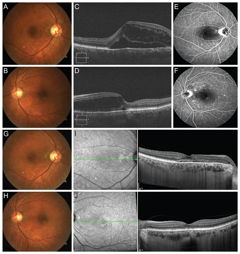
Fig. 1 At the initial visit, macular edema and serous detachment on both eyes were observed by fundus photo (A, right eye [OD]; B, left eye [OS]) and spectral domain optical coherence tomography (C, OD; D, OS). On fluorescence angiography, fluorescein dye pooling on the macula and multiple leakages were observed in both eyes (E, OD; F, OS). At 4 weeks after the initial visit, macular edema and serous detachment were much improved on fundus photo (G, OD; H, OS) and spectral domain optical coherence tomography (I, OD; J, OS).
Reference
-
1. Attal N, Cruccu G, Baron R, et al. EFNS guidelines on the pharmacological treatment of neuropathic pain: 2010 revision. Eur J Neurol. 2010; 17:1113–e88.2. Hilton EJ, Hosking SL, Betts T. The effect of antiepileptic drugs on visual performance. Seizure. 2004; 13:113–128.3. Parke-Davis MP. Product information: Neurontin (gabapentin). New York: Pfizer;2009.4. Herranz JL, Sol JM, Hernandez G. Gabapentin used in 559 patients with partial seizures: a multicenter observation study. Spanish Gabapentin Work Group. Rev Neurol. 2000; 30:1141–1145.5. Steinhoff BJ, Freudenthaler N, Paulus W. The influence of established and new antiepileptic drugs on visual perception. 1. A placebo-controlled, double-blind, single-dose study in healthy volunteers. Epilepsy Res. 1997; 29:35–47.
- Full Text Links
-
- Actions
-
Cited
- CITED
-
- Close
- Share
-
- Similar articles
-
- Risk Factors for Diffuse Diabetic Macular Edema as Classified by Optical Coherence Tomography
- The Effect of Intravitreal Triamcinolone Acetonide Injection according to the Diabetic Macular Edema Type
- The Correlation between Visual Acuity and Patterns of Diabetic Macular Edema in OCT Images
- Laser Photocoagulation in Diabetic Macular Edema
- Laser Photocoagulation in Diabetic Macular Edema
