Yonsei Med J.
2013 Jul;54(4):1049-1052. 10.3349/ymj.2013.54.4.1049.
Crown-Rump Length Measured in the Early First Trimester as a Predictor of Low Birth Weight
- Affiliations
-
- 1Department of Obstetrics and Gynecology, School of Medicine, Eulji University, Seoul, Korea. pwi3110@eulji.ac.kr
- 2Department of Preventive Medicine, School of Medicine, Eulji University, Seoul, Korea.
- KMID: 2158244
- DOI: http://doi.org/10.3349/ymj.2013.54.4.1049
Abstract
- The aim of this study is to assess the association between crown-rump length (CRL) measured before the 10th gestational week and birth weight. Results from 316 transvaginal ultrasonography scans at the 46th, 53rd, 60th, 67th, and 74th days of pregnancy were compared in low birth weight (LBW) versus normal birth weight groups. A positive correlation between CRL and birth weight was observed when CRL was measured at days 60, 67, and 74. CRL measured on the 67th day of pregnancy was significantly smaller in the LBW group than in the normal birth weight group. A cut-off value of CRL=26.5 mm measured at day 67 has the highest power to predict LBW.
Keyword
MeSH Terms
Figure
-
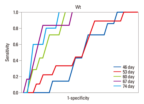
Fig. 1 ROC curve for prediction of low birth weight (<2500 g) for each ultrasonography date. Gestational days 60, 67 and 74 have larger areas under the curve than other days. ROC, receiver operating characteristic.
Reference
-
1. Kramer MS. Determinants of low birth weight: methodological assessment and meta-analysis. Bull World Health Organ. 1987; 65:663–737.2. Ligi I, Grandvuillemin I, Andres V, Dignat-George F, Simeoni U. Low birth weight infants and the developmental programming of hypertension: a focus on vascular factors. Semin Perinatol. 2010; 34:188–192.
Article3. Dickey RP, Gasser RF. Ultrasound evidence for variability in the size and development of normal human embryos before the tenth post-insemination week after assisted reproductive technologies. Hum Reprod. 1993; 8:331–337.
Article4. Smith GC, Smith MF, McNay MB, Fleming JE. First-trimester growth and the risk of low birth weight. N Engl J Med. 1998; 339:1817–1822.
Article5. Pardo J, Peled Y, Yogev Y, Melamed N, Ben-Haroush A. Association of crown-rump length at 11 to 14 weeks' gestation and risk of a large-for-gestational-age neonate. J Ultrasound Med. 2010; 29:1315–1319.
Article6. Thorsell M, Kaijser M, Almström H, Andolf E. Large fetal size in early pregnancy associated with macrosomia. Ultrasound Obstet Gynecol. 2010; 35:390–394.
Article7. Bukowski R, Smith GC, Malone FD, Ball RH, Nyberg DA, Comstock CH, et al. Fetal growth in early pregnancy and risk of delivering low birth weight infant: prospective cohort study. BMJ. 2007; 334:836.
Article8. Salomon LJ, Hourrier S, Fanchin R, Ville Y, Rozenberg P. Is first-trimester crown-rump length associated with birthweight? BJOG. 2011; 118:1223–1228.
Article9. Testart J, Frydman R. Minimum time lapse between luteinizing hormone surge or human chorionic gonadotropin administration and follicular rupture. Fertil Steril. 1982; 37:50–53.
Article10. Garcia JE, Jones GS, Wright GL Jr. Prediction of the time of ovulation. Fertil Steril. 1981; 36:308–315.
- Full Text Links
-
- Actions
-
Cited
- CITED
-
- Close
- Share
-
- Similar articles
-
- Transvaginal Measurements of Fetal Crown-Rump Length during the First Trimester after In Vitro Fertilization and Embryo Transfer in Korean Women
- Significance of fetal heart rate by vaginal ultrasound in the early pregnancy
- Comparison of Ultrasonographic Biometry and Regular Last Menstrual Period as Predictors of Day of Delivery in the Spontaneous Onset of Labor
- Maternal Weight Gain Pattern and Birth Weight
- Serum Ferritin Concentration in the Early Third Trimester of Pregnancy and Risk of Preterm Birth and Low Birth Weight Based on Gestational Age
