Malignant Fibrous Histiocytoma of Chest Wall
- Affiliations
-
- 1Department of Diagnostic Radiology, Yonsei University, College of Medicine, Research Institute of Radiological Science, Yonsei University, Seoul, Korea. kbrrdoh@yumc.yonsei.ac.kr
- KMID: 2158132
- DOI: http://doi.org/10.3349/ymj.2005.46.1.177
Abstract
- Primary malignant fibrous histiocytoma (MFH) of the chest wall is rare. We report a case of primary MFH arising from the chest wall, which was thought to be a metastasis or myeloma. The imaging study revealed a single mass of the chest wall involving a rib. Resection and chest wall reconstruction was done. The histologic diagnosis was storiform-pleomorphic primary MFH. Although MFH of the chest wall is an uncommon pathology, it should be considered in the differentiation of a single bony destructive lesion involving the rib with a soft tissue component.
MeSH Terms
Figure
-

Fig. 1 The posteroanterior (A) and lateral (B) chest radiograph revealed a round mass lesion (arrows) in the left anterior chest.
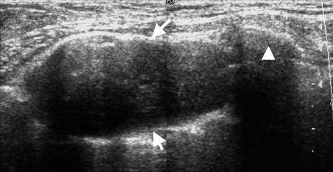
Fig. 2 Sonography showed a well defined, oval shaped, hypoechoic mass (arrows) involving a rib (arrowhead).

Fig. 3 CT demonstrated the presence of a relatively well defined, ovoid shaped mass at the anterior chest wall. A. Bony infiltration (arrow) and defect (arrowhead) was suggestive of the tumor having a malignant nature. The interface between the mass and the lung was smooth, and compression of the lung was observed. B. The mass showed heterogeneous weak enhancement (arrow). There was no mediastinal lymph node enlargement.
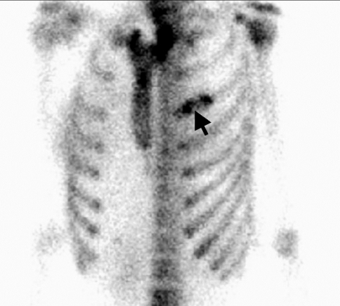
Fig. 4 The WBBS revealed markedly increased bony uptake (arrow) of the left 3rd anterior rib without other abnormal uptake on the whole skeletons.
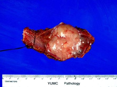
Fig. 5 Gross specimen showed a multilobulated pinkishwhite solid mass.

Fig. 6 Microscopic findings. A. The mass adhered to and infiltrated the inner surface of the rib (arrow). One segment of the mass (arrowhead) situated within the bony defect (H-E stain, × 40). B. Storiform arrangement of malignant cells, including a plump cytoplasm and a few mitotic figures, was suggestive of storiform-pleomorphic malignant fibrous histiocytoma (H-E stain, × 100).
Reference
-
1. Scott WW, Scott PP, Trerotola SO. Radiology of the thoracic skeleton. 1990. Philadelphia: BC Decker.2. Vock P. Higgins C, Pettersson H, editors. Magnetic resonance imaging and computed tomography of the chest wall. Chest and cardiac radiology. 1991. Vol 1. London: Merit Publishing International;p. 162–185. NICER series on diagnostic imaging.3. Belal A, Kandil A, Allam A, Khafaga Y, El-Husseiny G, El-Enbaby A, et al. Malignant fibrous histiocytoma: a retrospective study of 109 cases. Am J Clin Oncol. 2002; 25:16–22. PMID: 11823689.4. Sawai H, Kamiya A, Kurahashi S, Yamanaka Y, Manabe T. Malignant fibrous histiocytoma originating from the chest wall: report of a case and collective review of cases. Surg Today. 1998; 28:459–463. PMID: 9590721.
Article5. Enzinger FM, Weiss SW. Soft tissue tumors. 1995. 3rd ed. St. Louis: Mosby.6. Shinjo K. Analysis of prognostic factors and chemotherapy of malignant fibrous histiocytoma of soft tissue: a preliminary report. Jpn J Clin Oncol. 1994; 24:154–159. PMID: 8007425.
Article7. Spanier SS, Floyd J. A clinicopathologic comparison of malignant fibrous histiocytoma and liposarcoma. Instr Course Lect. 1989; 38:407–417. PMID: 2539413.8. Fukunaka H, Etoh T, Nakagawa H, Tamaki K. A case of subcutaneous malignant fibrous histiocytoma circumscribed by fibrous tissue. J Dermatol. 1996; 23:836–839. PMID: 8990710.
Article9. Kuhlman JE, Bouchardy L, Fishman EK, Zerhouni EA. CT and MR imaging evaluation of chest wall disorders. RadioGraphics. 1994; 14:571–595. PMID: 8066273.
Article10. Tateishi U, Kusumoto M, Hasegawa T, Yokoyama R, Moriyama N. Primary malignant fibrous histiocytoma of the chest wall: CT and MR appearance. J Comput Assist Tomogr. 2002; 26:558–563. PMID: 12218820.
Article11. Munk PL, Sallomi DF, Janzen DL, Lee MJ, Connell DG, O'Connell JX, et al. Malignant fibrous histiocytoma of soft tissue imaging with emphasis on MRI. J Comput Assist Tomogr. 1998; 22:819–826. PMID: 9754124.
Article12. Murphey MD, Gross TM, Rosenthal HG. From the archives of the AFIP. musculoskeletal malignant fibrous histiocytoma: radiologic-pathologic correlation. RadioGraphics. 1994; 14:807–826. PMID: 7938770.
Article13. Dahlin DC, Unni KK. Bone tumors: General Aspects and Data on 8,542 Cases. 1986. 4th ed. Springfied: Thomas.14. Bielack SS, Schroeders A, Fuchs N, Bacci G, Bauer HC, Mapeli S, et al. Malignant fibrous histiocytoma of bone: a retrospective EMSOS study of 125 cases. European Musculo-Skeletal Oncology Society. Acta Orthop Scand. 1999; 70:353–360. PMID: 10569265.15. Gibbs JF, Huang PP, Lee RJ, McGrath B, Brooks J, McKinley B, et al. Malignant fibrous histiocytoma: an institutional review. Cancer Invest. 2001; 19:23–27. PMID: 11291552.
- Full Text Links
-
- Actions
-
Cited
- CITED
-
- Close
- Share
-
- Similar articles
-
- Malignant fibrous histiocytoma
- A Case of the retroperitoneal Malignant Fibrous Histiocytoma
- Malignant Fibrous Histiocytoma: A Case Report
- Long-Term Survival after Wide Resection of Malignant Fibrous Histiocytoma of the Chest Wall
- Progression of Dermatofibrosarcoma Proruberans to Malignant Fibrous Histiocytoma: Report of a case
