Yonsei Med J.
2005 Feb;46(1):86-94. 10.3349/ymj.2005.46.1.86.
The Utility of Multi-detector Row Spiral CT for Detection of Coronary Artery Stenoses
- Affiliations
-
- 1Division of Cardiology, Cardiovascular Hospital, Yonsei University College of Medicine, Korea. ycchoi@yumc.yonsei.ac.kr
- 2Division of Radiology, Yonsei University, Seoul, Korea.
- KMID: 2158117
- DOI: http://doi.org/10.3349/ymj.2005.46.1.86
Abstract
- Contrast-enhanced multi-detector row spiral computed tomography (MDCT) was introduced as a promising noninvasive method for vascular imaging. This study examined the accuracy of this technique for detecting significant coronary artery stenoses. Both MDCT (Sensation 16, Siemens, Germany, 12 x 0.75 mm collimation and 0.42 sec rotation speed, 120 kV, 500 effective mA, and 2.7 mm/rotation table-feed) and invasive coronary angiography (CAG) were performed on 61 patients (mean age 59.2 +/- 10, 44 men) who were suspected of having coronary artery disease. All patients were treated with atenolol (25 - 50 mg) prior to imaging and the heart rate was maintained below 65 beats per minutes during image acquisition. The images were reconstructed in the diastole around TI - 400 ms with a 0.5 mm increment and a 1.0 mm thickness. All coronary arteries with a diameter of 2.0 mm or more were assessed for the presence of a stenosis (> 50% luminal narrowing). Two independent radiologists who were unaware of the results of the invasive CAG evaluated the MDCT data, and the results were compared with those from the invasive CAG (interval 1- 27, mean 11 days). An evaluation of the CT coronary angiogram (CTCA) was possible in 58 of the 61 patients (95%). Image acquisition of the major coronary arteries including the left main trunk was available in 229 out of 244 arteries. Invasive CAG showed that 35 out of 58 patients had significant coronary artery stenoses by. patient analysis of those who could be evaluated showed that CT coronary angiography correctly classified 30 out of 35 patients as having at least 1 coronary stenosis (sensitivity 85.7%, specificity 91.3%, positive predictive value 93.8%, negative predictive value 80.8%). By analyzing each coronary artery, CAG found 62 stenotic coronary arteries in the 229 coronary arteries that could be evaluated. MDCT correctly detected 50 out of 62 stenotic coronary arteries and an absence of stenosis was correctly identified in 156 out of 167 normal coronary arteries (sensitivity 80.6%, specificity 93.4%, positive predictive value 81.9%, negative predictive value 92.8%). The non-invasive technique of MDCT for examining the coronary artery appears to be a useful method for detecting coronary artery stenoses with a high accuracy particularly with the proximal portion and large arteries.
Keyword
MeSH Terms
Figure
-
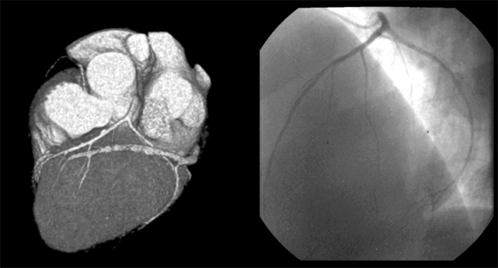
Fig. 1 Normal coronary artery in MDCT and CAG.

Fig. 2 Significant stenosis at proximal RCA.

Fig. 3 Detection of significant stenosis at the left main trunk by MDCT.

Fig. 4 Stent patency evaluation by MDCT. The stent patency evaluation was possible by a density measurement and a direct visual assessment in the lumen of the stent.
Cited by 1 articles
-
Off-Pump Coronary Artery Bypass Grafting in Moyamoya Disease
Do-Kyun Kim, Kyung-Jong Yoo
Yonsei Med J. 2007;48(5):876-878. doi: 10.3349/ymj.2007.48.5.876.
Reference
-
1. Fayad ZA, Fuster V. Clinical imaging of the high-risk or vulnerable atherosclerotic plaque. Circ Res. 2001. 89:305–316.2. Klingenbeck-Regn K, Schaller S, Flohr T, Ohnesorge B, Kopp AF, Baum U. Subsecond multi-slice computed tomography: basics and applications. Eur J Radiol. 1999. 31:110–124.3. Knez A, Becker CR, Ohnesorge B, Haberl R, Reiser M, Steinbeck G. Noninvasive detection of coronary artery stenosis by multislice helical computed tomography. Circulation. 2000. 101:221–222.4. Nieman K, Oudkerk M, Rensing BJ, Ooijen P, Munne A, Geuns RJ, et al. Coronary angiography with multislice computed tomography. Lancet. 2001. 357:599–603.5. Kopp AF, Schroeder S, Kuettner A, Baumbach A, Georg C, Kuzo R, et al. Non-invasive coronary angiography with high resolution multidetector-row computed tomography. Eur Heart J. 2002. 23:1714–1725.6. Achenbach S, Giesler T, Ropers D, Ulzheimer S, Derlien H, Schulte C, et al. Detection of coronary artery stenoses by contrast-enhanced, retrospectively ECG-gated, multi-slice spiral CT. Circulation. 2001. 103:2535–2538.7. Knez A, Becker CR, Leber A, Ohnesorge B, Becker A, White C, et al. Usefulness of multislice spiral computed tomography angiography for determination of coronary artery stenoses. Am J Cardiol. 2001. 88:1191–1194.8. Vogl TJ, Abolmaali ND, Diebold T, Engelmann K, Ay M, Dogan S, et al. Techniques for the detection of coronary atherosclerosis: multi-detector row CT coronary angiography. Radiology. 2002. 223:212–220.9. Schroeder S, Kopp AF, Kuettner A, Burgstahler C, Herdeg C, Heuschmid M, et al. Influence of heart rate on vessel visibility in noninvasive coronary angiography using new multislice computed tomography: experience in 94 patients. Clin Imaging. 2002. 26:106–111.10. Giesler T, Baum U, Ropers D, Ulzheimer S, Wenkel E, Mennicke M, et al. Noninvasive visualization of coronary arteries using contrast-enhanced multidetector CT: Influence of heart rate on image quality and stenosis detection. Am J Roentgenol. 2002. 179:911–916.11. Ropers D, Baum U, Pohle K, Anders K, Ulzheimer S, Ohnesorge B, et al. Detection of coronary artery stenoses with thin-slice multi-detector row spiral computed tomography and multiplanar reconstruction. Circulation. 2003. 107:664–666.12. Detre KM, Wright E, Murphy ML, Takaro T. Observer agreement in evaluating coronary angiograms. Circulation. 1975. 52:979–986.13. Becker CR, Ohnesorge BM, Joseph Schoepf U, Reiser MF. Current development of cardiac imaging with multi-detector row CT. Eur J Radiol. 2000. 36:97–103.14. Achenbach S, Moshage W, Ropers D, Nossen J, Daniel WG. Value of electron-beam computed tomography for the detection of high-grade coronary artery stenoses and occlusions. N Engl J Med. 1998. 339:1964–1971.15. Ha JW, Cho SY, Shim WH, Chung N, Jang Y, Lee HM, et al. Noninvasive evaluation of coronary artery bypass graft patency using three-dimensional angiography obtained with contrast-enhanced electron beam CT. Am J Roentgenol. 1999. 172:1055–1059.16. Nieman K, Cademartiri F, Lemos PA, Raaijmakers R, Pattynama PM, de Feyter PJ. Reliable noninvasive coronary angiography with fast submillimeter multislice spiral computed tomography. Circulation. 2002. 106:2051–2054.17. Reddy G, Chernoff DM, Adams JR, Higgins CB, et al. Coronary artery stenoses: assessment with contrast-enhanced electron-beam CT and axial reconstructions. Radiology. 1998. 208:167–172.18. Schmermund A, Rensing BJ, Sheedy PF, Bell MR, Rumberger JA. Intravenous electron-beam computed tomographic coronary angiography for segmental analysis of coronary artery stenoses. J Am Coll Cardiol. 1998. 31:1547–1554.19. Rumberger JA. Noninvasive coronary angiography using computed tomography Ready to kick it up another notch? Circulation. 2002. 106:2036–2038.20. Pump H, Mohlenkamp S, Sehnert CA, Schimpf SS, Schmidt A, Erbel R, et al. Coronary arterial stent patency: assessment with electron-beam CT. Radiology. 2000. 214:447–452.21. Mohlenkamps S, Pump H, Baumgart D, Haude M, Gronemeyer DH, Seibel RM, et al. Minimally invasive evaluation of coronary stents with electron beam computed tomography: in vivo and in vitro experience. Catheter Cardiovasc Interv. 1999. 48:39–47.22. Maintz D, Juergens KU, Wichter T, Grude M, Heindel W, Fischbach R. Imaging of coronary artery stents using multislice computed tomography: in vitro evaluation. Eur Radiol. 2003. 13:830–835.23. Fayad ZA, Fuster V, Nikolaou K, Becker C. Computed tomography and magnetic resonance imaging for noninvasive coronary angiography and plaque imaging: Current and potential future concepts. Circulation. 2002. 106:2026–2034.
- Full Text Links
-
- Actions
-
Cited
- CITED
-
- Close
- Share
-
- Similar articles
-
- Unusual Coronary Artery Fistula: Left Anterior Descending Coronary Artery - Left Ventricular Fistula Diagnosed by ECG-Gated Multi-Detector Row Coronary CT Angiography
- Diagnostic accuracy of 64-slice multi-detector CT coronary angiography in the evaluation of coronary artery disease
- A Case of Coronary Arteriovenous Fistula Associated with Pulmonary Artery Aneurysm Confirmed by Multi Detector-Row Helical CT
- MDCT Application in the Vascular System
- Detection of Hepatocelluar Carcinoma on Triple-Phase Images of Liver Using Multi-Detector Row Helical CT
