Progress in Malassezia Research in Korea
- Affiliations
-
- 1Department of Dermatology, Konkuk University School of Medicine, Seoul, Korea. kjahn@kuh.ac.kr
- KMID: 2157439
- DOI: http://doi.org/10.5021/ad.2015.27.6.647
Abstract
- Yeasts of the genus Malassezia are part of the normal flora of human skin. However, they are also associated with various skin diseases. Since the introduction of Malassezia to the Korean Dermatologic Society two decades ago, remarkable progress has been made in our knowledge of this genus. In this paper, we review recent developments in Malassezia research, including taxonomy and methods for species identification, recent genome analyses, Malassezia species distribution in healthy conditions and in specific skin diseases, trials investigating the mechanisms underlying Malassezia-related diseases, as well as therapeutic options. This review will enhance our understanding of Malassezia yeasts and related skin diseases in Korea.
Keyword
MeSH Terms
Figure
-
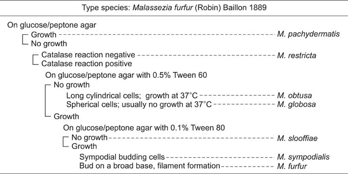
Fig. 1 Key to species of the genus Malassezia. Guého et al. (Antonie Van Leeuwenhoek 1996;69:337-355)12 and Ahn (Korean J Med Mycol 1998;3:81-88)2.
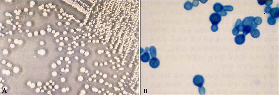
Fig. 2 Malassezia globosa. (A) Medium-sized, lighter in color, friable and crenated flat colonies with a pointed button center (Leeming and Notman medium, 34℃, 14 days). (B) Spherical, circular cells with buds on a narrow base (Parker Quink-KOH stain, ×1,000). Data from the article of Ahn (Korean J Med Mycol 1998;3:81-88)2.
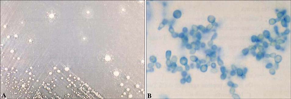
Fig. 3 Malassezia restrica. (A) Small-sized, circular, umbonate, entire, dull colonies (Leeming and Notman medium, 34℃, 14 days). (B) Small, spherical or oval cells with buds on a relatively narrow base (Parker Quink-KOH stain, ×1,000). Data from the article of Ahn (Korean J Med Mycol 1998;3:81-88)2.
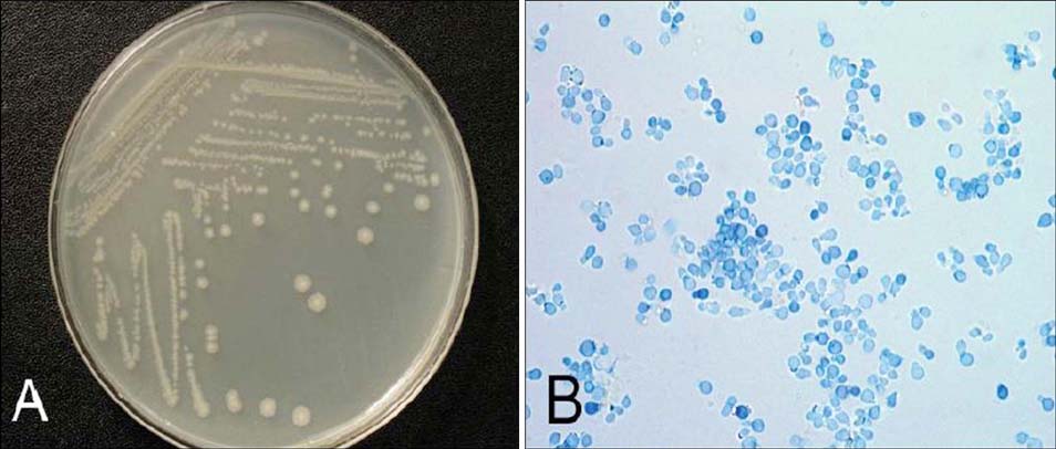
Fig. 4 Malassezia dermatis. (A) Large-sized, circular, smooth colonies (Leeming and Notman medium, 34℃, 14 days). (B) Spherical, oval, or ellipsoidal vegetative cells with monopolar budding (Parker Quink-KOH stain, ×1,000). Data from the article of Lim et al. (Korean J Dermatol 2007;45:1020-1030)52.
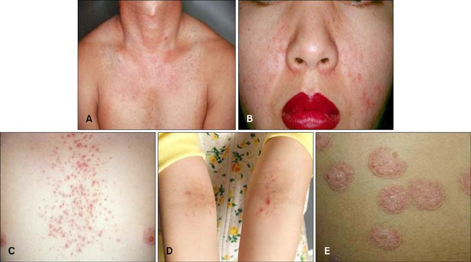
Fig. 5 Skin diseases associated with Malassezia species. (A) Pityriasis versicolor, (B) seborrheic dermatitis, (C) Malassezia folliculitis, (D) atopic dermatitis, and (E) psoriasis.
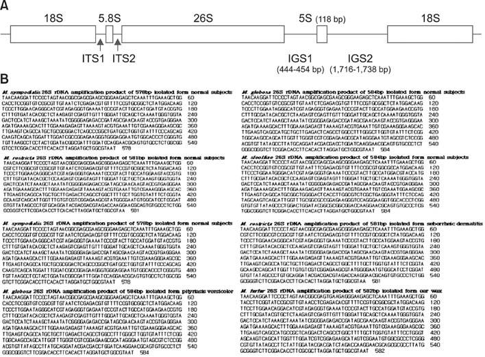
Fig. 6 (A) Schematic representation of the rRNA gene in the type strain (CBS 7966) of Malassezia globosa: the 26S rDNA, internal transcribed spacer (ITS1) region, and intergenic spacer 1 (IGS1) region sequences are used for species identification and strain typing. Data from the article of Sugita et al. (J Clin Microbiol 2003;41:3022-3027)48. (B) Sequences of the amplified 26S rDNA products from clinical isolates of Malassezia. Data from the article of Lee et al. (Korean J Med Mycol 2006;11:141-153)51.
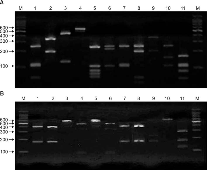
Fig. 7 Polymerase chain reaction (PCR) and restriction fragment length polymorphism patterns of the 26S rDNA PCR products digested with (A) Hha1 and (B) BstF51 from Malassezia standard strains. Lane M, 100-bp DNA ladder; Lane 1, Malassezia furfur; Lane 2, M. sympodialis; Lane 3, M. globosa; Lane 4, M. restricta; Lane 5, M. slooffiae; Lane 6, M. pachydermatis; Lane 7, M. japonica; Lane 8, M. nana; Lane 9, M. dermatis; Lane 10, M. obtusa; and Lane 11, M. yamatoensis. Data from the article of Lee et al. (Korean J Med Mycol 2006;11:141-153)51.
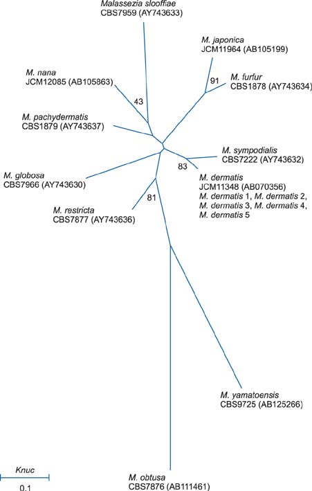
Fig. 8 A molecular phylogenetic tree constructed by the neighbor-joining method using the internal transcribed spacer 1 sequences of members of the genus Malassezia. Data from the article of Lim et al. (Korean J Dermatol 2007;45:1020-1030)52.
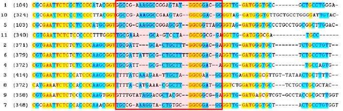
Fig. 9 Sequence alignment of the rRNA internal transcribed spacer 2 variable regions (box) of 11 Malassezia standard strains for pyrosequencing. Lane 1, Malassezia dermatis; Lane 2, M. furfur; Lane 3, M. globosa; Lane 4, M. japonica; Lane 5, M. nana; Lane 6, M. obtusa; Lane 7, M. pachydermatis; Lane 8, M. restricta; Lane 9, M. slooffiae; Lane 10, M. sympodialis; and Lane 11, M. yamatoensis. Data from the article of Song et al. (Korean J Med Mycol 2007;12:189-197)54.
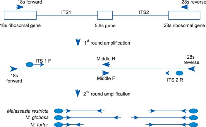
Fig. 10 Nested polymerase chain reaction: structure of the internal transcribed spacer (ITS) gene region and location of primer sites. Data from the articles of Lim et al. (Korean J Dermatol 2008;46:446-452)55.
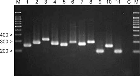
Fig. 11 Nested polymerase chain reaction products from standard Malassezia species. Lane M, molecular marker; Lane 1, Malassezia dermatis; Lane 2, M. furfur; Lane 3, M. globosa; Lane 4, M. japonica; Lane 5, M. nana; 6, M. obtusa; Lane 7, M. pachydermatis; Lane 8, M. restricta; Lane 9, M. slooffiae; Lane 10, M. sympodialis; Lane 11, M. yamatoensis; and Lane C, negative control. Data from the article of Lim et al. (Korean J Dermatol 2008;46:446-452)55.
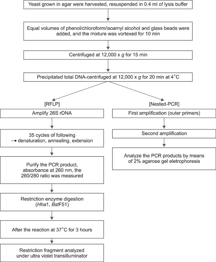
Fig. 12 Flowchart of the nested polymerase chain reaction (PCR) and restriction fragment length polymorphism (PCR-RFLP) methods. Data from the article of Oh et al. (Ann Dermatol 2009;21:352-357)58.
Reference
-
1. Kundu RV, Garg Amit. Yeast infections: Candidiasis, Tinea (Pityriasis) Versicolor, and Malassezia (Pityrosporum) Folliculitis. In : Goldsmith LA, Katz SI, Gilchrest BA, Paller AS, Leffell DJ, Wolff K, editors. Fitzpatrick's dermatology in general medicine. 8th ed. New York: McGraw-Hill;2012. p. 2298–2311.2. Ahn KJ. Taxonomy of the genus Malassezia. Korean J Med Mycol. 1998; 3:81–88.3. Gupta AK, Batra R, Bluhm R, Boekhout T, Dawson TL Jr. Skin diseases associated with Malassezia species. J Am Acad Dermatol. 2004; 51:785–798.
Article4. Kwon-Chung KJ, Bennett JE. Medical mycology. . Philadelphia: Lea & Febiger;1992. p. 170–182.5. Benham RW. The cultural characteristics of Pityrosporum ovale-a lipophilic fungus. J Invest Dermatol. 1939; 2:187–203.6. Ingham E, Cunningham AC. Malassezia furfur. J Med Vet Mycol. 1993; 31:265–288.
Article7. Dorn M, Roehnert K. Dimorphism of Pityrosporum orbiculare in a defined culture medium. J Invest Dermatol. 1977; 69:244–248.8. Porro MN, Passi S, Caprilli F, Mercantini R. Induction of hyphae in cultures of Pityrosporum by cholesterol and cholesterol esters. J Invest Dermatol. 1977; 69:531–534.
Article9. Cannon PF. International Commission on the Taxonomy of Fungi (ICTF): name changes in fungi of microbiological, industrial and medical importance. Part 2. Microbiol Sci. 1986; 3:285–287.10. Leeming JP, Notman FH. Improved methods for isolation and enumeration of Malassezia furfur from human skin. J Clin Microbiol. 1987; 25:2017–2019.
Article11. Cunningham AC, Leeming JP, Ingham E, Gowland G. Differentiation of three serovars of Malassezia furfur. J Appl Bacteriol. 1990; 68:439–446.12. Guého E, Midgley G, Guillot J. The genus Malassezia with description of four new species. Antonie Van Leeuwenhoek. 1996; 69:337–355.
Article13. Simmons RB, Guého E. A new species of Malassezia. Myco Res. 1990; 94:1146–1149.
Article14. Sugita T, Takashima M, Shinoda T, Suto H, Unno T, Tsuboi R, et al. New yeast species, Malassezia dermatis, isolated from patients with atopic dermatitis. J Clin Microbiol. 2002; 40:1363–1367.
Article15. Sugita T, Takashima M, Kodama M, Tsuboi R, Nishikawa A. Description of a new yeast species, Malassezia japonica, and its detection in patients with atopic dermatitis and healthy subjects. J Clin Microbiol. 2003; 41:4695–4699.
Article16. Sugita T, Tajima M, Takashima M, Amaya M, Saito M, Tsuboi R, et al. A new yeast, Malassezia yamatoensis, isolated from a patient with seborrheic dermatitis, and its distribution in patients and healthy subjects. Microbiol Immunol. 2004; 48:579–583.
Article17. Hirai A, Kano R, Makimura K, Duarte ER, Hamdan JS, Lachance MA, et al. Malassezia nana sp. nov., a novel lipid-dependent yeast species isolated from animals. Int J Syst Evol Microbiol. 2004; 54:623–627.
Article18. Cabañes FJ, Theelen B, Castellá G, Boekhout T. Two new lipid-dependent Malassezia species from domestic animals. FEMS Yeast Res. 2007; 7:1064–1076.19. Cabañes FJ, Vega S, Castellá G. Malassezia cuniculi sp. nov., a novel yeast species isolated from rabbit skin. Med Mycol. 2011; 49:40–48.
Article20. Ahn KJ, Ashbee HR. Determination of minimum inhibitory concentrations of several azole antifungals for Malassezia furfur. Ann Dermatol. 1996; 8:187–194.
Article21. Eichstedt E. Pilzbildung in der Pityriasis versicolor. Frorip Neue Notizen aus dem Gebeite der Naturkunde Heilkinde. 1846; 39:270–271.22. Ahn KJ. Malassezia species cultured from the lesions of pityriasis versicolor. Korean J Dermatol. 1997; 35:736–743.23. Yu HJ, Kim YS, Yang HY, Kim JH, Lee SK, Son SJ. The incidences of Malassezia in steroid acne and other acneiform eruptions. Korean J Med Mycol. 1998; 3:24–32.24. Hwang SJ, Lee SH, Kim DW, Jun JB, Chung SL, Jung HJ. Comparison of clinical manifestations and distribution of Malassezia species in patients with seborrheic dermatitis and atopic dermatitis. Korean J Dermatol. 1998; 36:617–627.25. Kang SH, Kim HU. The isolation of Malassezia yeasts in the comedones of acne vulgaris. Korean J Med Mycol. 1999; 4:33–39.26. Kim TY, Jang IG, Park YM, Kim HO, Kim CW. Head and neck dermatitis: the role of Malassezia furfur, topical steroid use and environmental factors in its causation. Clin Exp Dermatol. 1999; 24:226–231.
Article27. Oh CK, Kwon KS, Lee CW, Cho SH, Jang HS, Park JH. The distribution of Malassezia yeasts on the skin of neonates and infants. Korean J Med Mycol. 1999; 4:27–32.28. Ahn KJ, Kim KJ, Yi GJ. Efficacy of one-week regimen of itraconazole for pityriasis versicolor. Korean J Med Mycol. 1999; 4:124–130.29. Kwon HC, Kang SH, Kim HU. The distribution of Malassezia yeasts on normal human skin by culture study using the scrub-wash techique. Korean J Dermatol. 1999; 37:38–45.30. Kwon HC, Kang SH, Kim HU. The distibution of Malassezia yeasts on normal human skin by culture study using the swabbing technique. Korean J Dermatol. 1999; 37:46–56.31. Kim SC, Kim HU. The distribution of Malassezia species on the normal human skin according to body region. Korean J Med Mycol. 2000; 5:120–128.32. Lee YW, Kang HJ, Ahn KJ. Malassezia species cultured from the lesions of seborrheic dermatitis. Korean J Med Mycol. 2001; 6:70–76.33. Youn NH, Cha SH, Park SD. Malassezia yeasts in acne vulgaris. Korean J Dermatol. 2002; 40:1453–1460.34. Lee JH, Lee HS, Eun HC, Cho KH. Successful treatment of dandruff with 1.5% ciclopirox olamine shampoo in Korea. J Dermatolog Treat. 2003; 14:212–215.
Article35. Jang SJ, Choi YB, Ahn KJ. Malassezia species cultured from the lesions of Malassezia folliculitis. Korean J Med Mycol. 2003; 8:55–62.36. Choe YB, Jang SJ, Yim SM, Ahn KJ. The quantitative study on the distribution of Malassezia yeasts on the normal skin of the young adults. Korean J Med Mycol. 2004; 9:174–181.37. Jang SJ, Yim SM, Lee YW, Choe YB, Ahn KJ. The distribution of Malassezia yeasts on the earwax of young adults. Korean J Dermatol. 2005; 43:1583–1589.38. Lee YW, Yim SM, Lim SH, Choe YB, Ahn KJ. Quantitative investigation on the distribution of Malassezia species on healthy human skin in Korea. Mycoses. 2006; 49:405–410.
Article39. Moon HS, Son SJ, Park K, Chae JD. Difference of Malassezia species and Pityrosporum ovale specific IgE in head and neck lesions of atopic dermatitis related to ages and severity. Korean J Med Mycol. 2009; 14:1–8.40. Song HS, Kim SK, Kim YC. Comparison between Malassezia folliculitis and non-Malassezia folliculitis. Ann Dermatol. 2014; 26:598–602.41. Senczek D, Siesenop U, Böhm KH. Characterization of Malassezia species by means of phenotypic characteristics and detection of electrophoretic karyotypes by pulsed-field gel electrophoresis (PFGE). Mycoses. 1999; 42:409–414.42. Guillot J, Deville M, Berthelemy M, Provost F, Guého E. A single PCR-restriction endonuclease analysis for rapid identification of Malassezia species. Lett Appl Microbiol. 2000; 31:400–403.
Article43. Theelen B, Silvestri M, Guého E, van Belkum A, Boekhout T. Identification and typing of Malassezia yeasts using amplified fragment length polymorphism (AFLP), random amplified polymorphic DNA (RAPD) and denaturing gradient gel electrophoresis (DGGE). FEMS Yeast Res. 2001; 1:79–86.
Article44. Gaitanis G, Velegraki A, Frangoulis E, Mitroussia A, Tsigonia A, Tzimogianni A, et al. Identification of Malassezia species from patient skin scales by PCR-RFLP. Clin Microbiol Infect. 2002; 8:162–173.
Article45. Gupta AK, Kohli Y, Summerbell RC. Molecular differentiation of seven Malassezia species. J Clin Microbiol. 2000; 38:1869–1875.
Article46. Makimura K, Tamura Y, Kudo M, Uchida K, Saito H, Yamaguchi H. Species identification and strain typing of Malassezia species stock strains and clinical isolates based on the DNA sequences of nuclear ribosomal internal transcribed spacer 1 regions. J Med Microbiol. 2000; 49:29–35.
Article47. Sugita T, Suto H, Unno T, Tsuboi R, Ogawa H, Shinoda T, et al. Molecular analysis of Malassezia microflora on the skin of atopic dermatitis patients and healthy subjects. J Clin Microbiol. 2001; 39:3486–3490.
Article48. Sugita T, Kodama M, Saito M, Ito T, Kato Y, Tsuboi R, et al. Sequence diversity of the intergenic spacer region of the rRNA gene of Malassezia globosa colonizing the skin of patients with atopic dermatitis and healthy individuals. J Clin Microbiol. 2003; 41:3022–3027.
Article49. Sugita T, Tajima M, Tsubuku H, Tsuboi R, Nishikawa A. Quantitative analysis of cutaneous Malassezia in atopic dermatitis patients using real-time PCR. Microbiol Immunol. 2006; 50:549–552.
Article50. Tajima M, Sugita T, Nishikawa A, Tsuboi R. Molecular analysis of Malassezia microflora in seborrheic dermatitis patients: comparison with other diseases and healthy subjects. J Invest Dermatol. 2008; 128:345–351.
Article51. Lee YW, Lim SH, Ahn KJ. The application of 26S rDNA PCR-RFLP in the identification and classification of Malassezia yeast. Korean J Med Mycol. 2006; 11:141–153.52. Lim SH, Kim SM, Jung BR, Lee YW, Choe YB, Ahn KJ. A mycological and molecular biological study of Malassezia dermatis isolated from Korean. Korean J Dermatol. 2007; 45:1020–1030.53. Kim SM, Lim SH, Jung BR, Lee YW, Choe YB, Ahn KJ. The application of colony PCR in the molecular biological analysis of Malassezia yeasts. Korean J Med Mycol. 2007; 12:180–188.54. Song YC, Lim SH, Jung BR, Lee YW, Choe YB, Ahn KJ. The application of pyrosequencing method in the identification and classification of Malassezia yeasts. Korean J Med Mycol. 2007; 12:189–197.55. Lim SW, Shin MG, Lim JY, Yun SJ, Kim SJ, Lee SC, et al. Nested PCR for detection of Malassezia species from patient skin scales and clinical strains. Korean J Dermatol. 2008; 46:446–452.56. Lee YW, Kim SM, Oh BH, Lim SH, Choe YB, Ahn KJ. Isolation of 19 strains of Malassezia dermatis from healthy human skin in Korea. J Dermatol. 2008; 35:772–777.
Article57. Jang SJ, Lim SH, Ko JH, Oh BH, Kim SM, Song YC, et al. The investigation on the distribution of Malassezia yeasts on the normal Korean skin by 26S rDNA PCR-RFLP. Ann Dermatol. 2009; 21:18–26.
Article58. Oh BH, Song YC, Lee YW, Choe YB, Ahn KJ. Comparison of nested PCR and RFLP for identification and classification of Malassezia yeasts from Healthy human skin. Ann Dermatol. 2009; 21:352–357.
Article59. Kim SH, Ko HC, Kim MB, Kwon KS, Oh CK. The effect of detergents on the morphology and immunomodulatory activity of Malassezia furfur. Ann Dermatol. 2009; 21:130–135.
Article60. Yim SM, Kim JY, Ko JH, Lee YW, Choe YB, Ahn KJ. Molecular analysis of Malassezia microflora on the skin of the patients with atopic dermatitis. Ann Dermatol. 2010; 22:41–47.
Article61. Oh BH, Lee YW, Choe YB, Ahn KJ. Epidemiologic study of Malassezia yeasts in seborrheic dermatitis patients by the analysis of 26S rDNA PCR-RFLP. Ann Dermatol. 2010; 22:149–155.
Article62. Kim HM, Lim YY, Park EJ, Chun YJ, Kim MN, Kim BJ, et al. The development and evaluation of multiplex PCR technique for identification of Malassezia Yeast. Korean J Med Mycol. 2010; 15:51–60.63. Song YC, Hahn HJ, Kim JY, Ko JH, Lee YW, Choe YB, et al. Epidemiologic study of Malassezia yeasts in acne patients by analysis of 26S rDNA PCR-RFLP. Ann Dermatol. 2011; 23:321–328.
Article64. Lee YW, Byun HJ, Kim BJ, Kim DH, Lim YY, Lee JW, et al. Distribution of Malassezia species on the scalp in Korean seborrheic dermatitis patients. Ann Dermatol. 2011; 23:156–161.
Article65. Lim SH, Kim YR, Jung JW, Hahn HJ, Lee YW, Choe YB, et al. A comparison study between culture based technique and Op-site non-culture based technique for identifying Malassezia yeasts on normal skin. Korean J Med Mycol. 2012; 17:217–229.
Article66. DeAngelis YM, Saunders CW, Johnstone KR, Reeder NL, Coleman CG, Kaczvinsky JR Jr, et al. Isolation and expression of a Malassezia globosa lipase gene, LIP1. J Invest Dermatol. 2007; 127:2138–2146.67. Lee YW, Lee SY, Lee Y, Jung WH. Evaluation of expression of lipases and phospholipases of Malassezia restricta in patients with seborrheic dermatitis. Ann Dermatol. 2013; 25:310–314.
Article68. Kim JY, Hahn HJ, Choe YB, Lee YW, Ahn KJ, Moon KC. Molecular biological identification of Malassezia yeasts using pyrosequencing. Ann Dermatol. 2013; 25:73–79.
Article69. Lee WJ, Kim JY, Song CH, Jung HD, Lee SH, Lee SJ, et al. Disruption of barrier function in dermatophytosis and pityriasis versicolor. J Dermatol. 2011; 38:1049–1053.
Article70. Park HJ, Lee YW, Choe YB, Ahn KJ. Skin characteristics in patients with pityriasis versicolor using non-invasive method, MPA5. Ann Dermatol. 2012; 24:444–452.
Article71. Lee JW, Kim BJ, Kim MN. Photodynamic therapy: new treatment for recalcitrant Malassezia folliculitis. Lasers Surg Med. 2010; 42:192–196.
Article72. Wi HS, Na EY, Yun SJ, Lee JB. The antifungal effect of light emitting diode on Malassezia yeasts. J Dermatol Sci. 2012; 67:3–8.
Article73. Kim YR, Kim JH, Shin HJ, Choe YB, Ahn KJ, Lee YW. Clinical evaluation of a new-formula shampoo for scalp seborrheic dermatitis containing extract of Rosa centifolia petals and epigallocatechin gallate: a randomized, double-blind, controlled study. Ann Dermatol. 2014; 26:733–738.
Article
