Ann Dermatol.
2012 Feb;24(1):84-86. 10.5021/ad.2012.24.1.84.
An Acquired Anemic Patch Developed after a Cyst Excision: Is It a Variant of Nevus Anemicus?
- Affiliations
-
- 1Department of Dermatology and Cutaneous Biology Research Institute, Yonsei University College of Medicine, Seoul, Korea. oddung93@yuhs.ac
- 2Yeouido Oracle Cosmetic Dermatosurgery Clinic, Seoul, Korea.
- KMID: 2156863
- DOI: http://doi.org/10.5021/ad.2012.24.1.84
Abstract
- A 53-year-old male presented with a 6-year duration of a child's-palm sized hypopigmented patch located on his neck. He had a history of surgical excision of an epidermal cyst on the neck, and the hypopigmented patch developed about one month after the excision next to the surgery site. Application of cold or heat did not make the lesion distinct from the surrounding skin. Pressure on the lesion by a glass slide made the lesion indistinguishable from surrounding uninvolved lesions. Giving friction to the lesion failed to induce erythematous change, making it clearly visible. Histologically, the lesion showed normal findings with adequate numbers of melanocytes in the basal layer. Herein, we present an interesting case of an acquired anemic patch which developed after a cyst excision. We postulate that nerve damage after surgery that regulates the vascular tone of cutaneous vessels may have been an inducing event of the anemic patch in this patient.
Keyword
MeSH Terms
Figure
-
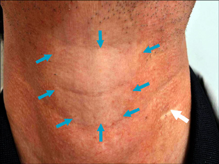
Fig. 1 A roughly child's-palm sized hypopigmented patch located on the neck (blue arrows) of the patient, and a 1.5 cm sized linear surgical scar (white arrow).
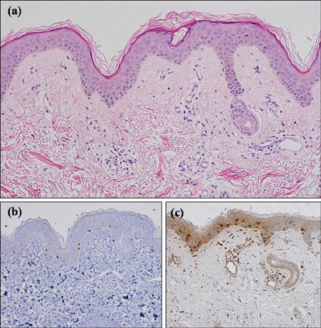
Fig. 2 histological findings by punch biopsy performed from the hypopigmented patch. (a) Normal findings with adequate numbers of melanocytes in the basal layer (H&E, ×100). Melan A (b) and S-100 protein (c) revealed a few melanocytes in the basal layer (×200).
Reference
-
1. Ahkami RN, Schwartz RA. Nevus anemicus. Dermatology. 1999. 198:327–329.
Article2. Miura Y, Tajima S, Ishibashi A, Hata Y. Multiple anemic macules on the arms: a variant form of nevus anemicus? Dermatology. 2000. 201:180–183.
Article3. Vörner H. Über Naevus anaemicus. Arch Dermatol Syphilis (Wien). 1906. 82:391–398.
Article4. Bruner E. Naevus anaemicus. Gaz Lek. 1912. 32:363–368.5. Daniel RH, Hubler WR, Wolf JE, Holder WR. Nevus anemicus. Donor-dominant defect. Arch Dermatol. 1977. 113:53–56.
Article6. Fleisher TL, Zeligman I. Nevus anemicus. Arch Dermatol. 1969. 100:750–755.
Article7. Mountcastle EA, Diestelmeier MR, Lupton GP. Nevus anemicus. J Am Acad Dermatol. 1986. 14:628–632.
Article8. Sarifakioglu E, Erdal E. Multiple anaemic macules of the arms: a variant of Bier's spots or naevus anemicus? J Eur Acad Dermatol Venereol. 2006. 20:892–893.
Article9. Grosshans E. Multiple anemic macules or Bier's spots? Dermatology. 2001. 202:272.
Article10. Tey HL. Approach to hypopigmentation disorders in adults. Clin Exp Dermatol. 2010. 35:829–834.
Article
- Full Text Links
-
- Actions
-
Cited
- CITED
-
- Close
- Share
-
- Similar articles
-
- Phakomatosis Pigmentovascularis
- Clinical Study of Nevus Anemicus
- Bilateral Ota Nevus and Bilateral Ito Nevus: 2 cases of extensive dermal melanocytic nevi associated with vascular nevus
- A Case of Congenital Melanocytic Nevus Combined with an Epidermal Cyst
- A Case of Acquired Bilateral Nevus of Ota-like Macules Accompanying the Common Blue Nevus
