Cutaneous Metaplastic Synovial Cyst of the First Metatarsal Head Area
- Affiliations
-
- 1Department of Dermatology, College of Medicine, Hallym University, Anyang, Korea. dermakkh@yahoo.co.kr
- 2Department of Dermatology, College of Medicine, Hallym University, Chuncheon, Korea.
- KMID: 2156780
- DOI: http://doi.org/10.5021/ad.2011.23.S2.S165
Abstract
- A cutaneous metaplastic synovial cyst (CMSC) is a cyst lined with metaplastic synovial tissue, which includes the formation of an intracystic villous structure resembling hyperplastic synovial villi. Clinically, the lesion is a tender, subcutaneous nodule that usually occurs at the site of previous surgical trauma and is frequently misdiagnosed as a suture granuloma. The actual cause remains unclear; however, trauma is presumed to be a precipitating factor, as most reported cases have demonstrated a history of antecedent cutaneous injury. Here, we present a case of CMSC in a 51-year-old woman who presented with a cystic mass localized in the left sole. She had no history of previous trauma or surgical procedures performed in the area. Although the case explained in this report is a spontaneous case of CMSC that occurred without a history of trauma, it is believed to have been caused by constant and chronic pressure since CMSC occurred in the first metatarsal head area, a part of the sole where heavy pressure is consistently applied.
MeSH Terms
Figure
-
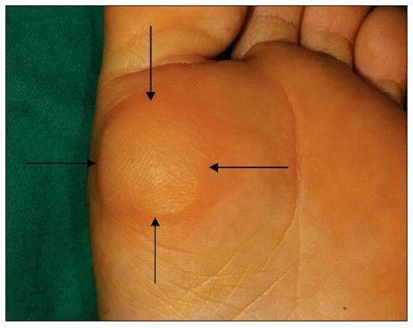
Fig. 1 Solitary, well-demarcated 3.0×3.0 cm-sized, skin-colored hyperkeratotic mass with cystic consistency.
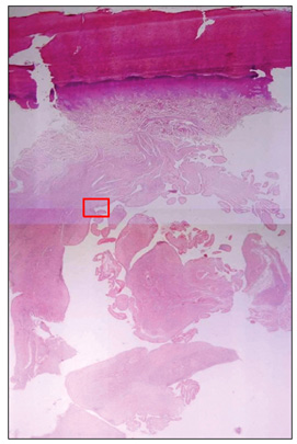
Fig. 2 Dermal cyst containing numerous villous-like structures that consisted of hyaline fibrous tissue projecting toward the center of cyst cavity (H&E, ×40).

Fig. 3 Magnification of the rectangle in Fig. 2. The villous structures were lined by membrane of varying cellularity, which mimicked synovium. Some were composed of hyalinized connective tissue covered with by fibrin whereas others were highly cellular and were lined by multilayers of epithelioid and fibroblastic cells (H&E, ×200).
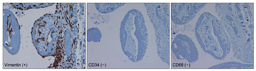
Fig. 4 Immunohistochemistry showed that the cells lining the cyst reacted positively for vimentin, but negative for CD34 and CD68 (×100).
Reference
-
1. Gonzalez JG, Chiselli RW, Santa Cruz DJ. Synovial metaplasia of the skin. Am J Surg Pathol. 1987. 11:343–350.
Article2. Bhawan J, Dayal Y, González-Serva A, Eisen R. Cutaneous metaplastic synovial cyst. J Cutan Pathol. 1990. 17:22–26.
Article3. Nieto S, Buezo GF, Jones-Caballero M, Fraga J. Cutaneous metaplastic synovial cyst in an Ehlers-Danlos patient. Am J Dermatopathol. 1997. 19:407–410.
Article4. Stern DR, Sexton FM. Metaplastic synovial cyst after partial excision of nevus sebaceous. Am J Dermatopathol. 1988. 10:531–535.5. Gómez Dorronsoro ML, Martinez-Peñuela JM, Ruiz de la Hermosa J. Metaplastic synovial cyst. Am J Surg Pathol. 1988. 12:649–650.6. Singh SR, Ma AS, Dixon A. Multiple cutaneous metaplastic synovial cysts. J Am Acad Dermatol. 1999. 41:330–332.
Article7. Lin YC, Tsai TF. Cutaneous metaplastic synovial cyst: unusual presentation with "a bag of worms". Dermatol Surg. 2003. 29:198–200.
Article8. Chakravarthy KM, Lavery KM, Barrett AW. Recurrent cutaneous metaplastic synovial cyst. Oral Surg Oral Med Oral Pathol Oral Radiol Endod. 2007. 103:e42–e44.
Article9. Choonhakarn C, Tang S. Cutaneous metaplastic synovial cyst. J Dermatol. 2003. 30:480–484.
Article10. Goiriz R, Ríos-Buceta L, Alonso-Pérez A, Jones-Caballero M, Fraga J, García-Diez A. Cutaneous metaplastic synovial cyst. J Am Acad Dermatol. 2005. 53:180–181.
Article11. Ramdial PK, Singh Y, Singh B. Metaplastic synovial cyst in male breast. Ann Diagn Pathol. 2005. 9:219–222.
Article12. Guala A, Viglio S, Ottinetti A, Angeli G, Canova G, Colombo E, et al. Cutaneous metaplastic synovial cyst in Ehlers-Danlos syndrome: report of a second case. Am J Dermatopathol. 2008. 30:59–61.
Article13. Fujisawa Y, Ito M, Nakamura Y, Furuta J, Ishii Y, Kawachi Y, et al. Perforated ischiogluteal bursitis mimicking a gluteal decubitus ulcer in patients with spinal cord injury: report of 2 cases. Arch Dermatol. 2010. 146:932–934.
Article
- Full Text Links
-
- Actions
-
Cited
- CITED
-
- Close
- Share
-
- Similar articles
-
- Cutaneous Metaplastic Synovial Cyst on the Finger after Orthopedic Surgery
- Cutaneous Metaplastic Synovial Cyst of the Cheek Generated by Repetitive Minor Trauma
- Synovial Chondromatosis of the First Metatarsal(A Case Report)
- Synovial Chondromatosis: Report of 4 cases
- Lumbar Radiculopathy Caused by Intraspinal Synovial Cyst: A Case Report
