Large Dermal Non Neural Granular Cell Tumor on the Surgical Wound Site
- Affiliations
-
- 1Department of Dermatology, College of Medicine, Dankook University, Cheonan, Korea. 4exodus@hanmail.net
- KMID: 2156776
- DOI: http://doi.org/10.5021/ad.2011.23.S2.S147
Abstract
- Granular cell tumors (GCTs) can be divided into neural type with S-100 reactivity and non-neural type without that. The latter has not been widely recognized and there are only fewer reports available when compared to conventional GCT. A 65-year-old man was presented with the presence of a painless mass on his back. The mass had developed into a small nodule on the scar developed because of previous surgery carried out 2 years ago. The tumor consisted of large, polygonal cells comprising of an enormous number of faintly eosinophilic small granules in the cytoplasm. The cytoplasmic granules were stained positively for periodic acid-Schiff stain. Immunohistochemical stains for S-100 protein and neuron-specific enolase were found to be negative. Herein, we report the appearance of a very rare case of non neural GCT developed on the surgical scar in support with relevant literature reviews.
Keyword
MeSH Terms
Figure
-
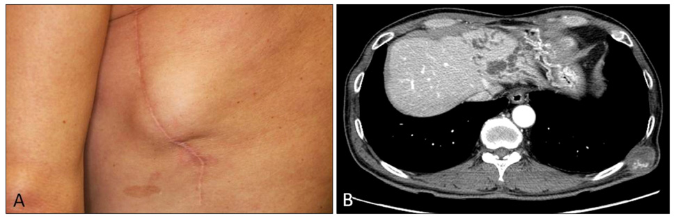
Fig. 1 (A) Solitary hard, fixed dermal mass on the left lateral chest wall. (B) Computed tomography scan revealing the presence of an oval mass with dimensions of 3.6×2.5 cm in the subcutaneous layer adjacent to intercostal muscle.
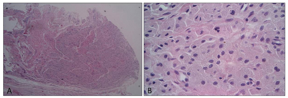
Fig. 2 (A) Diffuse infiltration of monomorphic tumor cells on the dermis (H&E, ×40). (B) Large, polygonal tumor cells with abundant faintly eosinophilic granular cytoplasm (H&E, ×400).
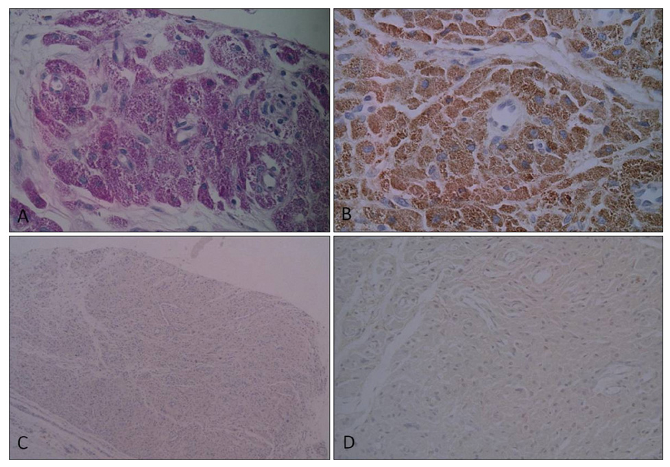
Fig. 3 (A) The cytoplasmic granules showing positive stain with periodic acid-Schiff (×400). (B) Immunohistochemical stains revealed positivity for CD68 (×400). (C, D) Stains for S-100 protein, neuron-specific enolase were negative (C: S-100, ×40, D: NSE, ×200).
Cited by 1 articles
-
Congenital Non-Neural Granular Cell Tumor Mimicking Nevus Lipomatosus Superficialis
Jun Yeong Park, Won Joo Kwon, Bok Won Park, Eun Byul Cho, Eun Joo Park, Kwang Ho Kim, Kwang Joong Kim
Ann Dermatol. 2017;29(6):776-778. doi: 10.5021/ad.2017.29.6.776.
Reference
-
1. Lazar AJ, Fletcher CD. Primitive nonneural granular cell tumors of skin: clinicopathologic analysis of 13 cases. Am J Surg Pathol. 2005. 29:927–934.2. LeBoit PE, Barr RJ, Burall S, Metcalf JS, Yen TS, Wick MR. Primitive polypoid granular-cell tumor and other cutaneous granular-cell neoplasms of apparent nonneural origin. Am J Surg Pathol. 1991. 15:48–58.
Article3. Murcia JM, Idoate M, Laparte C, Baldonado C. Granular cell tumor of vulva on episiotomy scar. Gynecol Oncol. 1994. 53:248–250.
Article4. Chaudhry IH, Calonje E. Dermal non-neural granular cell tumour (so-called primitive polypoid granular cell tumour): a distinctive entity further delineated in a clinicopathological study of 11 cases. Histopathology. 2005. 47:179–185.
Article5. Yeh I, Tran DT, Davis TL, Argenyi ZB. An infiltrative variant of non-neural granular cell tumor: a case report. J Cutan Pathol. 2009. 36:Suppl 1. 46–51.
Article6. Habeeb AA, Salama S. Primitive nonneural granular cell tumor (so-called atypical polypoidgranular cell tumor). Report of 2 cases with immunohistochemical and ultrastructural correlation. Am J Dermatopathol. 2008. 30:156–159.
Article7. Lee MW, Choi JH, Sung KJ, Moon KC, Koh JK. Granular cell tumor: clinical and histopathological study. Korean J Dermatol. 2000. 38:1030–1036.8. Sobel HJ, Avrin E, Marquet E, Schwarz R. Reactive granular cells in sites of trauma. A cytochemical and ultrastructural study. Am J Clin Pathol. 1974. 61:223–234.
Article9. Giménez-Bascuñana A, Piqueras-Pérez FM. Granular cell traumatic neuroma of salivary gland. Arch Pathol Lab Med. 2001. 125:1000–1001.
Article
