Ann Dermatol.
2008 Mar;20(1):18-21. 10.5021/ad.2008.20.1.18.
A Case of Melanoacanthoma: Immunohistochemical Staining Using VECTOR(R) NovaRED(TM) to Distinguish Melanocytes from the Cutaneous Pigment
- Affiliations
-
- 1Department of Dermatology, College of Medicine, Korea University, Seoul, Korea. choh@korea.ac.kr
- 2Department of Pathology, College of Medicine, Korea University, Seoul, Korea.
- KMID: 2156354
- DOI: http://doi.org/10.5021/ad.2008.20.1.18
Abstract
- Melanoacanthoma is a rare benign mixed tumor of both keratinocytes and melanocytes. Although some authors said that it is a rare variant of seborrheic keratosis, it has clinical and histological features distinct from seborrheic keratosis. It has large dendritic melanin-laden melanocytes throughout all levels of epidermis showing a disruption of melanin transfer from the melanocytes to neighboring keratinocytes. However, it is difficult to distinguish melanocytes clearly from cutaneous pigment in immunohistochemical stain with usually used brown chromogen. We used chromogen with brick-red indicator product (VECTOR(R) NovaRED(TM)) in S-100 and melan-A immunohistochemical staining to distinguish melanocytes from melanin laden keratinocytes. We suggest that the immunohistochemical staining using this novel chromogen may be useful in the diagnosis of melanoacanthoma.
MeSH Terms
Figure
-
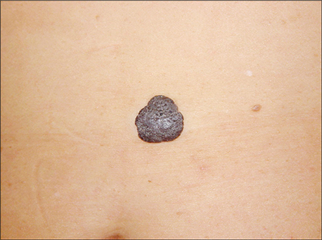
Fig. 1 About 2 × 2 cm-sized, well-demarcated, black-pigmented plaque on the abdomen.
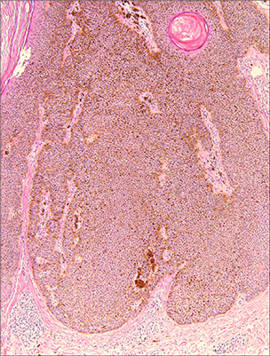
Fig. 2 The epidermis showed hyperkeratosis, acanthosis, papillomatosis and pseudo-horn cysts and the tumor was composed of basaloid and squamous keratinocytes and melanocytes (H&E, × 100).
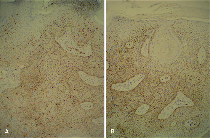
Fig. 3 The tumor consisted of a proliferation of keratinocytes with a low pigment content and numerous heavily stained large dendritic melanocytes spread throughout all levels of the stratum malpighii, which are stained red by chromogen VECTOR® NovaRED™ (A: S-100, × 100; B: melan-A, × 100).
Reference
-
1. Mishima Y, Pinkus H. Benign mixed tumor of melanocytes and malpighian cells. Melanoacanthoma: its relationship to bloch's benign non-nevoid melanoepithelioma. Arch Dermatol. 1960; 81:539–550.
Article2. Prince C, Mehregan AH, Hashimoto K. Large melanoacanthomas: a report of five cases. J Cutan Pathol. 1984; 11:309–317.
Article3. Vion B, Merot Y. Melanoacanthoma of the penis shaft. Report of a case. Dermatologica Basel. 1989; 179:87–89.4. Sexton FM, Maize JC. Melanotic macules and melanoacanthomas of the lip. A comparative study with census of the basal melanocyte population. Am J Dermatopathol. 1987; 9:438–444.5. Kihiczak GG, Centurion SA, Schwartz RA, Lambert WC. Giant cutaneous melanoacanthoma. Int J Dermatol. 2004; 43:936–937.
Article6. Simon P, Requena L, Sanchez Yus E. How rare is melanoacanthoma? Arch Dermatol. 1991; 127:583–584.
Article7. Schlappner OLA, Rowden G, Philips TM, Rahim Z. Melanoacanthoma: ultrastructural and immunological studies. J Cutan Pathol. 1978; 5:127–141.
Article8. Shidham VB, Qi DY, Acker S, Kampalath B, Chang CC, George V, et al. Evaluation of micrometastases in sentinel lymph nodes of cutaneous melanoma. Higher diagnostic accuracy with melan-A and MART-1 compared with S-100 protein and HMB-45. Am J Surg Pathol. 2001; 25:1039–1046.
Article9. Mouton RW, Jordaan HF, Schneider JW. A new type of minocycline-induced cutaneous hyperpigmentation. Clin Exp Dermatol. 2003; 29:8–14.
Article10. Lee GS, Ahn KJ, Kim JM, Lee ES. A histopathologic study of the seborrheic keratosis. Korean J Dermatol. 1992; 30:76–80.11. Kim JM, Kim JS, Cha MH, Lee CJ. A case of melanoacanthoma. Korean J Dermatol. 1984; 22:435–438.
