Angiomatoid Spitz Nevus
- Affiliations
-
- 1Department of Dermatology, College of Medicine, Dong-A University, Busan, Korea. khkim@dau.ac.kr
- KMID: 2156353
- DOI: http://doi.org/10.5021/ad.2008.20.1.14
Abstract
- Spitz nevus is a variant of melanocytic nevus which is histopathologically defined as large spindle and/or epithelioid cells. Angiomatoid Spitz nevus is a rare histologic variant of desmoplastic Spitz nevus characterized by prominent vasculature. We present a case of angiomatoid Spitz nevus, celluar type, that has not been reported before. We provide another example to show the remarkable diversity of Spitz nevus.
Figure
-

Fig. 1 A 0.8 × 0.8 × 0.6 cm pink, slightly pigmented dome-shaped papule on the auricle of the right ear.

Fig. 2 There was epidermal thinning with a partial epidermal collarette and tumor cells involving the full thickness of the dermis without epidermal involvement (A-inset) (H&E, original magnification × 10). Numerous small and large sized blood vessels are present in the upper and deep portion of the dermis (A) (H&E, original magnification × 200). Many spindle and epithelioid cells with abundant melanin granules are embedded in a desmoplastic stroma (B) (H&E, original magnification × 40). Small sized thick walled blood vessels are present in dermis (C) (H&E, original magnification × 400).
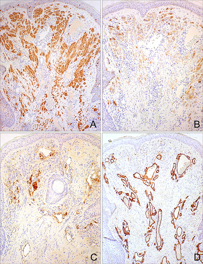
Fig. 3 Immunostaining for S-100 protein showed a positive reaction within the nevus cells (A), HMB-45 staining was partially positive within upper dermal nests (B), Factor VIII and CD34 was positive within blood vessels, respectively (C, D) (original magnification × 100).
Reference
-
1. Diaz-Cascajo C, Borghi S, Weyers W. Angiomatoid Spitz nevus: a distinct variant of desmoplastic spitz nevus with prominent vasculature. Am J Dermatopathol. 2000; 22:135–139.2. McKee PH, Calonje E, Granter SR. Spitz nevus, atypical spitz nevus and spitzoid melanoma. In : McKee PH, editor. Pathology of the skin. 3rd ed. London: Elsevier Mosby;2005. p. 1268–1275.3. Paniago-Pereira C, Maize JC, Ackerman AB. Nevus of large spindle and/or epithelioid cells (spitz's nevus). Arch Dermatol. 1978; 14:1811–1823.
Article4. Tomizawa K. Desmoplastic spitz nevus showing vascular proliferation more prominently in the deep portion. Am J Dermatopathol. 2002; 24:184–185.
Article5. Casso EM, Grin-Jorgensen CM, Grant-Kels JM. Spitz nevi. J Am Acad Dermatol. 1992; 27:901–913.
Article6. Jose RM, Bennett A, Holmes J. Spitz naevi presenting as pyogenic granulomata. Br J Plast Surg. 2005; 58:1037–1039.
Article7. Jang HS, Cha JH, Oh CK, Kwon KS. Spitz naevus showing clinical features of both granuloma pyogenicum and pigmented naevus. Br J Dermatol. 2001; 145:349–350.
Article
- Full Text Links
-
- Actions
-
Cited
- CITED
-
- Close
- Share
-
- Similar articles
-
- A Case of Angiomatoid Spitz Nevus with High Cellularity and Lymphovascular Tumor Emboli-like Features
- The first case of vaginal angiomatoid Spitz nevus causing vaginal bleeding
- Spitz Nevus with Atypical Clinical Features in a Baby
- Spitz Nevus in a Giant Speckled Lentiginous Nevus
- A Case of Speckled Lentiginous Nevus combined with Spitz Nevi
