Two Pilosebaceous Cysts with Apocrine Hidrocystoma in One Biopsy Site: A Spectrum of the Same Disease Process?
- Affiliations
-
- 1Department of Dermatology Inje University School of Medicine, Busan, Korea. drskin99@hanmail.net
- KMID: 2156352
- DOI: http://doi.org/10.5021/ad.2008.20.1.11
Abstract
- A 28-year-old woman presented with multiple, asymptomatic, erythematous to bluish papules located on the chest. Histopathologically, three round, well defined cystic structures were seen on the upper and lower dermis. The first cyst was milia, the second was apocrine hidrocystoma and the other, largest cyst was an eruptive vellus hair cyst (EVHC). A diagnosis of multiple pilosebaceous cysts combined with apocrine hidrocystoma was made. Since the milia and EVHC originate from the pilosebaceous unit, and the apocrine duct opens to the pilosebaceous orifice, we suggest that they can occur simultaneously in the same unit.
Figure
-
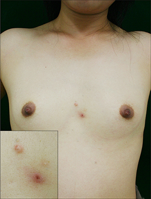
Fig. 1 Smooth surfaced, slightly erythematous, bluish papules on the chest; inset: close up view of the papule.

Fig. 2 (A) Histologic examination of the lesion showed round, well defined multiple cystic structures on the upper and lower dermis (H&E, × 12.5). (B) The upper cyst is composed of several layers of squamous and granular layer and filled with horny material arranged in laminated layers (H&E, × 200). (C) The cyst wall is lined by secretory cells showing decapitation secretion (H&E, × 200). (D) The cyst wall is lined by stratified squamous cell epithelium and the lumen contains vellus hair shaft (H&E, × 200).
Cited by 2 articles
-
A Case of Eruptive Vellus Hair Cysts That Developed on the Labium Major
Ju Hyuk Park, Young Her, Bo Mi Chun, Chul Woo Kim, Sang Seok Kim
Ann Dermatol. 2009;21(3):294-296. doi: 10.5021/ad.2009.21.3.294.Hypomelanosis of Ito with Multiple Congenital Anomalies
Da-Ae Yu, Ohsang Kwon, Kyu Han Kim
Ann Dermatol. 2019;31(5):576-580. doi: 10.5021/ad.2019.31.5.576.
Reference
-
1. Requena L, Sanchez-Yus E. Follicular hybrid cysts. An expanded spectrum. Am J Dermatopathol. 1991; 13:228–233.2. Iacobelli D, Hashimoto K, Kato I, Ito M, Suzuki Y. Clobetasol induced milia. J Am Acad Dermatol. 1989; 21:215–217.3. Weedon D. Cysts, sinuses and pits. Skin pathology. 2nd ed. London: Churchill Livingstone;2002. p. 508.4. Sanchez-Yus E, Aguilar-Martinez A, Cristobal-Gil MC, Urbina-Gonzalez F, Guerra-Rodriguez P. Eruptive vellus hair cyst and steatocystoma multiplex: two related conditions? J Cutan Pathol. 1988; 15:40–42.
Article5. Patrizi A, Neri I, Guerrini V, Costa AM, Passarini B. Persistent milia, steatocystoma multiplex and eruptive vellus hair cyst: variable expression of multiple pilosebaceous cysts within an affected family. Dermatology. 1998; 196:392–396.
Article6. Kurban SR, Bhawan J. Cutaneous cysts lined by nonsquamous epithelium. Am J Dermatopathol. 1991; 13:228–233.
Article7. Anderson WK, Rao BK, Bhawan J. The hybrid epidermoid and apocrine cyst. Am J Dermatopathol. 1996; 18:364–366.
Article8. Takeda H, Miura A, Katagata Y, Mitsuhashi Y, Kondo S. Hybrid cyst: case reports and review of 15 cases in Japan. J Eur Acad Dermatol Venereol. 2003; 17:83–86.
Article
