A Case of Otogenic Subdural Empyema That Progressed to Cerebellar Abscess after Radical Mastoidectomy
- Affiliations
-
- 1Department of Neurosurgery, College of Medicine, Hallym University, Chuncheon, Korea. ns1287@hallym.or.kr
- 2Department of Otorhinolaryngology-Head & Neck Surgery, College of Medicine, Hallym University, Chuncheon, Korea.
- KMID: 2156160
- DOI: http://doi.org/10.13004/jknts.2008.4.2.105
Abstract
- Nowadays, despite great advances in the treatment of all forms of infectious otitis media, otogenic complications still occur. Of all forms, complications in the central nervous system from otitis media represent the most life-threatening conditions, requiring immediate diagnosis and precise therapeutic intervention. Subdural empyema is a rare intracranial complication after otogenic procedures. We have experienced a case of otogenic subdural empyema that progressed to a cerebellar abscess after a radical mastoidectomy which was performed without drainage of the subdural empyema. We present this case with a review of the relevant literature.
Keyword
Figure
-
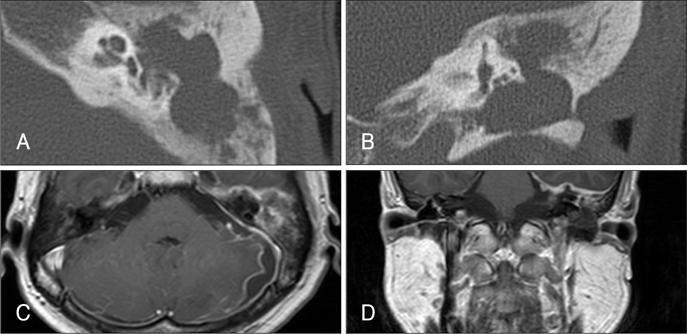
FIGURE 1 The findings of initial temporal bone CT and brain MRI. A: Temproal bone CT axial image shows soft tissue density filling left middle ear cavity and mastoid antrum. B: Temproal bone CT coronal image shows the bony stenosis of left external auditory canal. C: Enhanced T1-weighted MRI axial image shows subdural collection of low-signal intensity with peripheral rim enhancement, diffuse thickened enhancement in left posterior dura mater and leptomeningeal enhancement in left temporal lobe and cerebellar hemisphere. D: Enhanced T1-weighted MRI coronal image shows diffuse thickened enhancement in dura mater of left temporal lobe and focal dural defect suspiciously. CT: computerized tomography, MRI: magnetic resonance image.
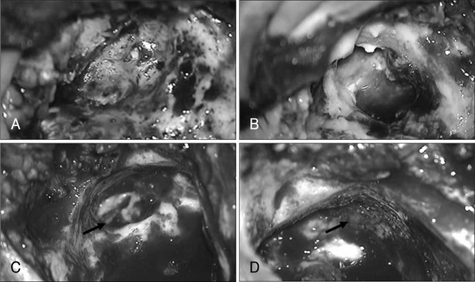
FIGURE 2 Intraoperative findings of radical mastoidectomy. A: The photo shows the stenosis of left external auditory canal. B: The photo shows auto-mastoidectomy state by cholesteatoma. C: The photo shows the fistula of promontory (arrow). D: The photo shows the exposure of dura mater by destruction of tegmen tympani. The dura mater of middle cerebral fossa is covered with adhesive granulation tissue (arrow).

FIGURE 3 Follow-up enhanced T1-weighted images in 2 weeks after surgery. The photos show multiple ring-like enhanced cavitary lesions in left cerebellar hemisphere, which are aggravated compared with initial study, and brain edema due to these lesions.
Reference
-
1. Bannister G, Williams B, Smith S. Treatment of subdural empyema. J Neurosurg. 1981; 55:82–88.
Article2. Barriere SL, Latwick LI, Jacobs RA, Conte JE. Vancomycin treatment for Enterococcal meningitis. Arch Neurol. 1985; 42:686–688.
Article3. Chung JW, Lee SW, Song HM, Kim JH. Otogenic brain abscess: recent experience of 13 cases. Korean J Otolaryngol-Head Neck Surg. 2004; 47:1054–1059.4. Greenlee JE. Subdural Empyema. Curr Treat Options Neurol. 2003; 5:13–22.
Article5. Hoyt DJ, Fisher SR. Otolaryngologic management of patients with subdural empyema. Laryngoscope. 1991; 101:20–24.
Article6. Kim KS, Jeon YS, Yang TY, Shin JS. A case of 'silent' subdural abscess detected incidentally during the operation of chronic otitis media with cholesteatoma. Korean J Otolaryngol-Head Neck Surg. 2003; 46:602–605.7. Matthews PC, Conlon CP, Berendt AR, Kayley J, Jefferies L, Atkins BL, et al. Outpatient parenteral antimicrobial therapy (OPAT): is it safe for selected patients to self-administer at home? A retrospective analysis of a large cohort over 13 years. J Antimicrob Chemother. 2007; 60:356–362.
Article8. Nathoo N, Nadvi SS, Gouws E, van Dellen JR. Craniotomy improves outcomes for cranial subdural empyemas: computed tomography-era experience with 699 patients. Neurosurgery. 2001; 49:872–877. discussion 877-878.
Article9. Polyzoidis KS, Vranos G, Exarchakos G, Argyropoulou MI, Korantzopoulos P, Skevas A. Subdural empyema and cerebellar abscess due to chronic otitis media. Int J Clin Pract. 2004; 58:214–217.
Article10. Saah D, Elidan J, Gomori M. Intracranial complications of otitis media. Ann Otol Rhinol Laryngol. 1997; 106:873–874.
Article11. Seven H, Coskun BU, Calis AB, Saylin I, Turgut S. Intracranial abscesses associated with chronic suppurative otitis media. Eur Arch Otorhinolaryngol. 2005; 262:847–851.
Article12. Tice AD, Strait K, Ramey R, Hoaglund PA. Outpatient parenteral antimicrobial therapy for central nervous system infections. Clin Infect Dis. 1999; 29:1394–1399.
Article13. Wackym PA, Canalis RF, Feurerman T. Subdural empyema of otorhinological origin. J Laryngol Otol. 1990; 104:118–122.
Article14. Zimmerman RA, Bilaniuk LR, Sze G. Intracranial infection. Brant-Zawadzki M, Norman D, editors. Magnetic resonance imaging of the central nervous system. 1987. New York: Raven;p. 235–257.
- Full Text Links
-
- Actions
-
Cited
- CITED
-
- Close
- Share
-
- Similar articles
-
- Subtentorial Empyema: Report of 2 Cases
- A Case of `Silent' Subdural Abscess Detected Incidentally during the Operation of Chronic Otitis Media with Cholesteatoma
- A Case of Subtentorial Subdural Empyema Resulting from Chronic Otitis Media with Cholesteatoma
- A Case of Acute Coalescent Mastoiditis with Otogenic Cerebellar Abscess due to Acute Otitis Media
- Two Cases of Cerebellar Abscess
