Anomalous External Carotid Artery-Internal Carotid Artery Anastomosis in Two Patients with Proximal Internal Carotid Arterial Remnants
- Affiliations
-
- 1Department of Neurology, Stroke Center, Myongji Hospital, Goyang 412-270, Korea.
- 2Department of Radiology, Seoul National University Hospital, Seoul National University College of Medicine, Seoul 110-744, Korea. aronnn@naver.com
- 3Department of Neurosurgery, Seoul National University Hospital, Seoul National University College of Medicine, Seoul 110-744, Korea.
- 4Department of Radiology, Asan Medical Center, University of Ulsan College of Medicine, Seoul 138-736, Korea.
- 5Department of Neurosurgery, Hallym University Sacred Heart Hospital, Hallym University College of Medicine, Anyang 431-796, Korea.
- KMID: 2155567
- DOI: http://doi.org/10.3348/kjr.2015.16.4.914
Abstract
- Two angiographic instances of anomalous external carotid artery (ECA) and internal carotid artery (ICA) anastomosis are described, each occurring at the C2-3 level and bearing remnants of proximal ICA. The ICA remnant of one patient (identifiable immediately upon bifurcation of the common carotid artery) was hypoplastic, and that of the other patient was an occluded arterial stump. These features are not typical of non-bifurcating ICA. The occipital artery originated from an anomalous connection in one instance and from the main trunk of the ECA (just past the ECA-ICA connection) in the other.
MeSH Terms
Figure
-
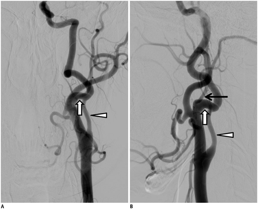
Fig. 1 44-year-old man with anomalous ECA-ICA anastomosis and hypoplastic proximal ICA. A, B. Frontal (A) and lateral (B) angiographic images showing anomalous ECA-ICA anastomosis at C2-3 spinal level and relatively hypoplastic proximal ICA at expected site of ICA origin. Anomalous anastomosis (white arrow), hypoplastic proximal ICA (arrowhead), and occipital artery (black arrow). ECA = external carotid artery, ICA = internal carotid artery
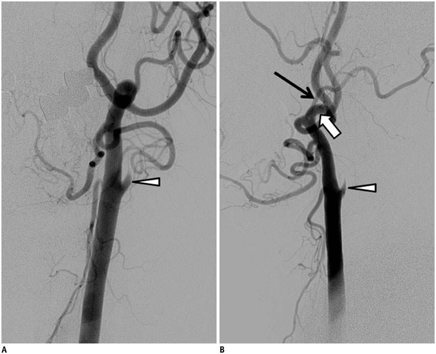
Fig. 2 60-year-old man with anomalous ECA-ICA anastomosis and proximal ICA remnant. A, B. Oblique (A) and lateral (B) angiographic images showing origins of ECA and ICA from common trunk, with carotid budding at carotid bifurcation level as probable remnant of proximal ICA. Anomalous anastomosis (white arrow), arterial stump (arrowhead), and occipital artery (black arrow). ECA = external carotid artery, ICA = internal carotid artery
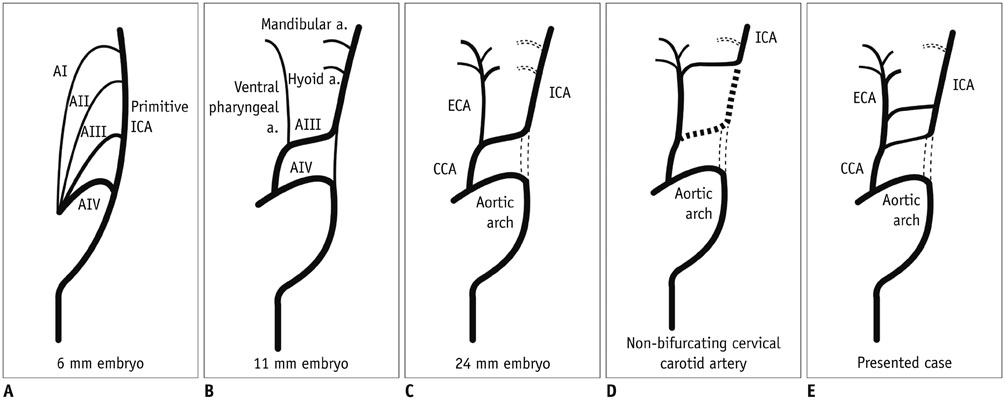
Fig. 3 Schematic illustration of presented case in comparison with normal ICA-ECA development and non-bifurcating cervical carotid artery. A-C. Illustration for development of normal internal carotid artery and external carotid artery. D, E. Illustrations for typical non-bifurcating cervical carotid artery (D) and presented cases (E). a = artery, AI = first aortic arch, AII = second aortic arch, AIII = third aortic arch, AIV = forth aortic arch, CCA = common carotid artery, ECA = external carotid artery, ICA = internal carotid artery
Reference
-
1. Ooigawa H, Nawashiro H, Fukui S, Tsuzuki N, Katoh H, Kawaguchi T, et al. Non-bifurcating cervical carotid artery. J Clin Neurosci. 2006; 13:944–947.2. Morimoto T, Nitta K, Kazekawa K, Hashizume K. The anomaly of a non-bifurcating cervical carotid artery. Case report. J Neurosurg. 1990; 72:130–132.3. Kaneko K, Akita M, Murata E, Imai M, Sowa K. Unilateral anomalous left common carotid artery; a case report. Ann Anat. 1996; 178:477–480.4. Nakai K, Kaji T, Uchino A, Kawauchi T, Tamura C, Otani N, et al. Congenital external carotid-internal carotid artery anastomosis associated with contralateral non-bifurcating cervical carotid artery. Neuroradiology. 2012; 54:521–523.5. Rodriguez HE, Ziauddin MF, Podbielski FJ, Durham JR, Clark ET. Congenital absence of the external carotid artery: atherosclerosis without a bifurcation. J Vasc Surg. 2002; 35:573–575.6. Klosek SK, Rungruang T. Topography of carotid bifurcation: considerations for neck examination. Surg Radiol Anat. 2008; 30:383–387.7. Suzuki T, Moriyama T, Moriwaki H, Yagihashi A, Yajima N, Takahashi G. Anomalous artery directly connecting the external and internal carotid arteries. Ann Anat. 2000; 182:59–63.8. Trainor PA, Krumlauf R. Hox genes, neural crest cells and branchial arch patterning. Curr Opin Cell Biol. 2001; 13:698–705.9. Kameda Y, Watari-Goshima N, Nishimaki T, Chisaka O. Disruption of the Hoxa3 homeobox gene results in anomalies of the carotid artery system and the arterial baroreceptors. Cell Tissue Res. 2003; 311:343–352.10. Chess MA, Barsotti JB, Chang JK, Ketonen LM, Westesson PL. Duplication of the extracranial internal carotid artery. AJNR Am J Neuroradiol. 1995; 16:1545–1547.11. Hacein-Bey L, Raghavan N, Mukundan G, Sekhon AK, Dodrill LK, Park TC. Fenestration of the petrous internal carotid artery with short segment duplication mimicking an arterial dissection. Clin Radiol. 2013; 68:972–975.
- Full Text Links
-
- Actions
-
Cited
- CITED
-
- Close
- Share
-
- Similar articles
-
- Congenital External Carotid-Internal Carotid Artery Anastomosis
- Anomalous Origin of the Vertebral Artery From the Internal Carotid Artery
- Angiographic Features of Unilateral Nonbifurcating Cervical Carotid Artery: A Case Report
- Patent Internal Carotid Artery with Collateral Circulation from Contralateral External Carotid Artery after Common Carotid Artery Occlusion
- Angiographic Demonstration of External Carotid-Vertebral Arteries Anastomosis
