Inter-Observer Variation in Ultrasound Measurement of the Volume and Diameter of Thyroid Nodules
- Affiliations
-
- 1Department of Radiology and Research Institute of Radiology, Asan Medical Center, University of Ulsan College of Medicine, Seoul 138-736, Korea. radbaek@naver.com
- KMID: 2155525
- DOI: http://doi.org/10.3348/kjr.2015.16.3.560
Abstract
OBJECTIVE
Thyroid nodule measurement using ultrasonography (US) is widely performed in various clinical scenarios. The purpose of this study was to evaluate inter-observer variation in US measurement of the volume and maximum diameter of thyroid nodules.
MATERIALS AND METHODS
This retrospective study included 73 consecutive patients with 85 well-defined thyroid nodules greater than 1 cm in their maximum diameter. US examinations were independently performed by using standardized measurement methods, conducted by two clinically experienced thyroid radiologists. The maximum nodule diameter and nodule volume, calculated from nodule diameters using the ellipsoid formula, were obtained by each reader. Inter-observer variations in volume and maximum diameter were determined using 95% Bland-Altman limits of agreement. The degree of inter-observer variations in volumes and the maximum diameters were compared using the Student's t test, between nodules < 2 cm in maximum diameter and those with > or = 2 cm.
RESULTS
The mean inter-observer difference in measuring the nodule volume was -1.6%, in terms of percentage of the nodule volume, and the 95% limit of agreement was +/- 13.1%. For maximum nodule diameter, the mean inter-observer difference was -0.6%, in terms of percentage of the nodule diameter, and the 95% limit of agreement was +/- 7.3%. Inter-observer variation in volume was greater in nodules of < 2 cm in maximum diameter, compared to the larger nodules (p = 0.035). However, no statistically significant difference was noted between the two groups regarding maximum nodule diameters (p = 0.511).
CONCLUSION
Any differences smaller than 13.1% and 7.3% in volume and maximum diameter, respectively, measured by using US for well-defined thyroid nodules of > 1 cm should not be considered as a real change in size.
MeSH Terms
Figure
-
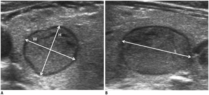
Fig. 1 Thyroid nodule diameters measured by using ultrasonography (US) is shown. On transverse US image (A), width (W) and height (H) of thyroid nodule were measured to determine maximum transverse diameter. Length (L) of thyroid nodule was considered as maximum diameter seen on longitudinal US image (B).
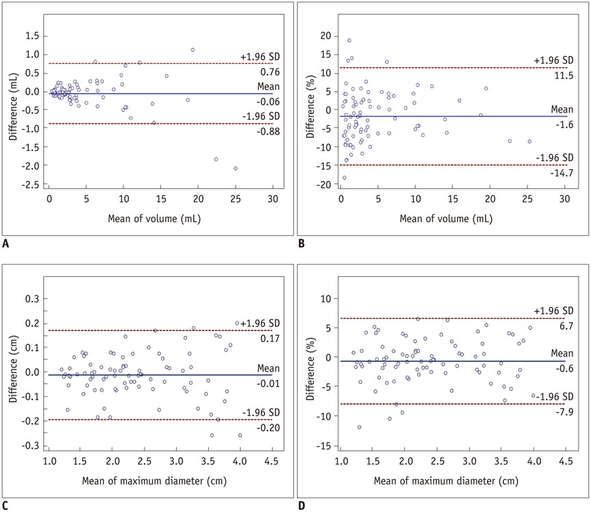
Fig. 2 Bland-Altman plots of interobserver agreement for thyroid nodule volume and maximum diameter. Bland-Altman plots of thyroid nodule volume (A, B) and maximum diameter (C, D) with measurement data (A, C) and standardized data (i.e., measurement differences as % of nodule volume and maximum diameter) (B, D) show relationship between two observers. Difference (y-axis) between two observers is plotted against mean value (x-axis) of two readers' measurements. Solid line indicates mean difference. Top and bottom dashed lines correspond to upper and lower margins of 95% limits of agreement. With probability of 95%, differences in normalized scores of future examinations will be between upper and lower limits of agreement (mean ± variability estimate = 1.96 standard deviation [SD]).
Cited by 2 articles
-
A Comparison of Ultrasound-Guided Fine Needle Aspiration versus Core Needle Biopsy for Thyroid Nodules: Pain, Tolerability, and Complications
Eun Ji Jeong, Sae Rom Chung, Jung Hwan Baek, Young Jun Choi, Jae Kyun Kim, Jeong Hyun Lee
Endocrinol Metab. 2018;33(1):114-120. doi: 10.3803/EnM.2018.33.1.114.Risk Factors for Tumor Size Increase During Active Surveillance of Papillary Thyroid Cancer: Meta-Analysis and Systematic Review
Doh Young Lee, Pilkeun Jang
Korean J Otorhinolaryngol-Head Neck Surg. 2021;64(12):914-921. doi: 10.3342/kjorl-hns.2021.00990.
Reference
-
1. American Thyroid Association (ATA) Guidelines Taskforce on Thyroid Nodules and Differentiated Thyroid Cancer. Cooper DS, Doherty GM, Haugen BR, Kloos RT, Lee SL, et al. Revised American Thyroid Association management guidelines for patients with thyroid nodules and differentiated thyroid cancer. Thyroid. 2009; 19:1167–1214.2. Gharib H, Papini E, Paschke R, Duick DS, Valcavi R, Hegedüs L, et al. American Association of Clinical Endocrinologists, Associazione Medici Endocrinologi, and EuropeanThyroid Association Medical Guidelines for Clinical Practice for the Diagnosis and Management of Thyroid Nodules. Endocr Pract. 2010; 16:Suppl 1. 1–43.3. Moon WJ, Baek JH, Jung SL, Kim DW, Kim EK, Kim JY, et al. Ultrasonography and the ultrasound-based management of thyroid nodules: consensus statement and recommendations. Korean J Radiol. 2011; 12:1–14.4. Baek JH, Kim YS, Lee D, Huh JY, Lee JH. Benign predominantly solid thyroid nodules: prospective study of efficacy of sonographically guided radiofrequency ablation versus control condition. AJR Am J Roentgenol. 2010; 194:1137–1142.5. Baek JH, Moon WJ, Kim YS, Lee JH, Lee D. Radiofrequency ablation for the treatment of autonomously functioning thyroid nodules. World J Surg. 2009; 33:1971–1977.6. Jeong WK, Baek JH, Rhim H, Kim YS, Kwak MS, Jeong HJ, et al. Radiofrequency ablation of benign thyroid nodules: safety and imaging follow-up in 236 patients. Eur Radiol. 2008; 18:1244–1250.7. Sung JY, Baek JH, Kim KS, Lee D, Yoo H, Kim JK, et al. Single-session treatment of benign cystic thyroid nodules with ethanol versus radiofrequency ablation: a prospective randomized study. Radiology. 2013; 269:293–300.8. Levine RA. Current guidelines for the management of thyroid nodules. Endocr Pract. 2012; 18:596–599.9. Hegedüs L, Karstrup S, Rasmussen N. Evidence of cyclic alterations of thyroid size during the menstrual cycle in healthy women. Am J Obstet Gynecol. 1986; 155:142–145.10. Bennedbaek FN, Nielsen LK, Hegedüs L. Effect of percutaneous ethanol injection therapy versus suppressive doses of L-thyroxine on benign solitary solid cold thyroid nodules: a randomized trial. J Clin Endocrinol Metab. 1998; 83:830–835.11. Døssing H, Bennedbaek FN, Hegedüs L. Effect of ultrasound-guided interstitial laser photocoagulation on benign solitary solid cold thyroid nodules - a randomised study. Eur J Endocrinol. 2005; 152:341–345.12. Brauer VF, Eder P, Miehle K, Wiesner TD, Hasenclever H, Paschke R. Interobserver variation for ultrasound determination of thyroid nodule volumes. Thyroid. 2005; 15:1169–1175.13. Moon WJ, Jung SL, Lee JH, Na DG, Baek JH, Lee YH, et al. Benign and malignant thyroid nodules: US differentiation--multicenter retrospective study. Radiology. 2008; 247:762–770.14. Frates MC, Benson CB, Charboneau JW, Cibas ES, Clark OH, Coleman BG, et al. Management of thyroid nodules detected at US: Society of Radiologists in Ultrasound consensus conference statement. Radiology. 2005; 237:794–800.
- Full Text Links
-
- Actions
-
Cited
- CITED
-
- Close
- Share
-
- Similar articles
-
- Thyroid Ultrasound: Change of Inter-observer Variability and Diagnostic Performance after Training
- Inter-observer and intra-observer reliability between manual segmentation and semi-automated segmentation for carotid vessel wall volume measurements on three-dimensional ultrasonography
- Volume Measurement Using 3-Dimensional US
- A comparison of three methods of assessing inter-observer variation applied to measurement of the symphysis-fundal height
- Proper Measurement of the Prostate Volume by Transrectal Ultrasound: Experimental Study about the Prostate with Focal Intravesical Protrusion of the Enlarged Central Gland
