Primary Hepatic Mucosa-Associated Lymphoid Tissue Lymphoma: A Case Report
- Affiliations
-
- 1Department of Radiology, Daejin Medical Center Bundang Jesaeng General Hospital, Seongnam, Korea. neverendlove@hanmail.net
- 2Department of Pathology, Daejin Medical Center Bundang Jesaeng General Hospital, Seongnam, Korea.
- 3Department of Internal Medicine, Daejin Medical Center Bundang Jesaeng General Hospital, Seongnam, Korea.
- KMID: 2155279
- DOI: http://doi.org/10.3348/jksr.2016.74.3.189
Abstract
- Primary hepatic mucosa-associated lymphoid tissue (MALT) lymphoma is an extremely rare lesion. Primary hepatic lymphomas are known to present as a single mass in > 70% of cases, and in many instances with no specific features on imaging. Herein, we described a case of primary hepatic MALT lymphoma in a 71-year-old woman. A computed tomography (CT) scan revealed a mass, 4.5 x 3.0 cm, in liver segment 2 (S2) that was poorly defined, with subtle enhancement during the arterial phase. Gadoxetic acid-enhanced magnetic resonance imaging also showed an arterially enhancing mass in S2, with low signal intensity during the hepatobiliary phase and high signal intensity on diffusion-weighted imaging with a high b-value. On fluorodeoxyglucose positron emission tomography/CT imaging, the mass showed a high standardized uptake value. Ultrasonography (US) revealed a hypoechoic mass, and US-guided core needle biopsy confirmed a hepatic MALT lymphoma.
MeSH Terms
Figure
-
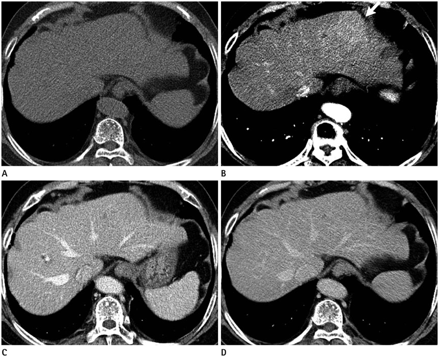
Fig. 1 Hepatic MALT lymphoma in a 71-year-old woman on CT. On unenhanced (A) and dynamic liver CT, the mass shows a mildly arterial enhancing mass (arrow) in liver segment 2 (B) in the arterial phase and iso-attenuation relative to the liver on unenhanced image (A), in the portal phase (C), and in the delayed phase (D). CT = computed tomography, MALT = mucosa-associated lymphoid tissue
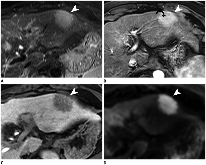
Fig. 2 Hepatic MALT lymphoma in a 71-year-old woman on gadoxetic acid-enhanced MRI. A. T2-weighted axial MR image shows a homogeneous high signal instensity mass (arrowhead) in liver segment 2. B, C. On gadoxetic acid-enhanced arterial phase (B), the mass shows homogeneous enhancement (arrowhead) and undistorted vessels traversing the mass (curved black arrow). The mass shows low signal intensity (arrowhead) in the hepatobiliary phase (C). D. With diffusion-weighted imaging, the intensity is high (arrowhead), with a high b-value (800 s/mm2). MALT = mucosa-associated lymphoid tissue, MRI = magnetic resonance imaging
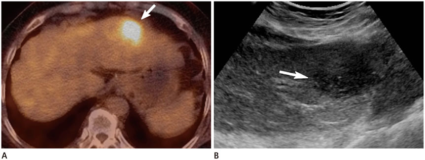
Fig. 3 Hepatic MALT lymphoma in a 71-year-old woman on FDG PET/CT and US. A. FDG PET/CT scanning reveals a high standardized uptake value (max SUV = 6.59) (arrow) at liver segment 2, suggesting a hypermetabolic mass. B. US demonstrates a hypoechoic solid mass (arrow) in liver segment 2. FDG PET/CT = fluorodeoxyglucose positron emission tomography computed tomography, MALT = mucosa-associated lymphoid tissue, US = ultrasonography

Fig. 4 Histopathologic findings and immunohistochemical staining of hepatic MALT lymphoma in a 71-year-old woman. A. The microscopic finding of the tumor includes a diffuse infiltration of small atypical lymphocytes expanding portal tracts, extending into lobules, and effacing normal hepatocytes (hematoxylin and eosin stain, × 100). B. Immunohistochemical staining reveals CD20-positive lymphoid cells (× 400). MALT = mucosa-associated lymphoid tissue
Reference
-
1. Matasar MJ, Zelenetz AD. Overview of lymphoma diagnosis and management. Radiol Clin North Am. 2008; 46:175–198, vii.2. Isaacson P, Wright DH. Malignant lymphoma of mucosa-associated lymphoid tissue. A distinctive type of B-cell lymphoma. Cancer. 1983; 52:1410–1416.3. Park JY, Choi MS, Lim YS, Park JW, Kim SU, Min YW, et al. Clinical features, image findings, and prognosis of inflammatory pseudotumor of the liver: a multicenter experience of 45 cases. Gut Liver. 2014; 8:58–63.4. Doi H, Horiike N, Hiraoka A, Koizumi Y, Yamamoto Y, Hasebe A, et al. Primary hepatic marginal zone B cell lymphoma of mucosa-associated lymphoid tissue type: case report and review of the literature. Int J Hematol. 2008; 88:418–423.5. Grazioli L, Olivetti L, Mazza G, Bondioni MP. MR imaging of hepatocellular adenomas and differential diagnosis dilemma. Int J Hepatol. 2013; 2013:374170.6. Apicella PL, Mirowitz SA, Weinreb JC. Extension of vessels through hepatic neoplasms: MR and CT findings. Radiology. 1994; 191:135–136.7. Jaffe ES. Malignant lymphomas: pathology of hepatic involvement. Semin Liver Dis. 1987; 7:257–268.8. Rodallec M, Guermazi A, Brice P, Attal P, Zagdanski AM, Frija J, et al. Imaging of MALT lymphomas. Eur Radiol. 2002; 12:348–356.9. Shiozawa K, Watanabe M, Ikehara T, Matsukiyo Y, Kikuchi Y, Kaneko H, et al. A case of contiguous primary hepatic marginal zone B-cell lymphoma and hemangioma ultimately diagnosed using contrast-enhanced ultrasonography. Case Rep Oncol. 2015; 8:50–56.
- Full Text Links
-
- Actions
-
Cited
- CITED
-
- Close
- Share
-
- Similar articles
-
- Longlasting Remission of Primary Hepatic Mucosa-associated Lymphoid Tissue (MALT) Lymphoma Achieved by Radiotherapy Alone
- Mucosa-Associated Lymphoid Tissue Lymphoma of the Esophagus Coexistent with Bronchus-Associated Lymphoid Tissue Lymphoma of the Lung
- Primary Mucosa-Associated Lymphoid Tissue Lymphoma of the Breast with Synchronous Contralateral Invasive Breast Cancer: A Case Report
- A case report of the Pulmonary Malignant Lymphomaof the mucosa-associated lymphoid tissue(MALT)
- Mucosa-Associated Lymphoid Tissue Lymphoma Arising from the Nasal Mucosa: A Case Report and Review of the Literature
