The Diagnostic Performance of Acoustic Radiation Force Impulse Elasticity Imaging to Differentiate Malignant from Benign Thyroid Nodules: Comparison with Conventional B-Mode Sonographic Findings
- Affiliations
-
- 1Department of Radiology, St. Vincent's Hospital, The Catholic University of Korea, Suwon, Korea. yparkh@catholic.ac.kr
- KMID: 2152601
- DOI: http://doi.org/10.3348/jksr.2016.74.2.96
Abstract
- PURPOSE
The purpose of this study was to evaluate the diagnostic performance of acoustic radiation force impulse (ARFI) elasticity imaging.
MATERIALS AND METHODS
One hundred and twenty-seven thyroid nodules were examined by both ARFI elastography and B-mode sonography. Virtual Touch tissue quantification (VTQ) values of the thyroid nodules were measured. Scoring of B-mode sonographic findings of each thyroid nodules was performed. The sums of these VTQ and the B-mode scores were determined. The comparative diagnostic performances of the VTQ value, the B-mode score, and the combined score were analyzed.
RESULTS
The sensitivity, specificity, positive predictive value, negative predictive value, and accuracy of each scoring mode were: B-mode score, 84%, 85%, 66%, 94%, and 85%; VTQ, 75%, 91%, 73%, 92%, and 86%; and combined score, 88%, 87%, 70%, 95%, and 88%. The areas under the curves for B-mode, VTQ, and combined score were 0.895, 0.837, and 0.912, respectively. Pairwise comparisons of receiver-operating characteristic curves showed no statistical differences between B-mode and VTQ, and B-mode and combined score. Combined score showed better diagnostic performance than VTQ value (p = 0.0023).
CONCLUSION
ARFI VTQ value is a good diagnostic modality for differentiating malignant thyroid nodules from benign nodules. However, ARFI evaluation is not superior to B-mode sonographic evaluation, but only has a better diagnostic performance when combined with B-mode sonographic findings.
MeSH Terms
Figure
-
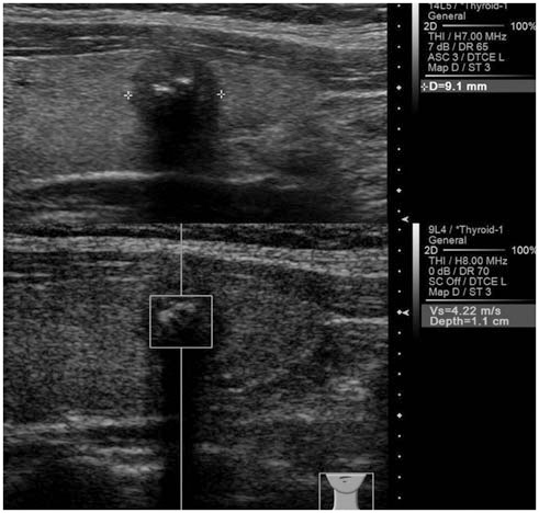
Fig. 1 Solid, marked hypoechoic nodule with calcification shows high VTQ score (4.22 m/s). This nodule was confirmed as papillary thyroid carcinoma. VTQ = Virtual Touch tissue quantification
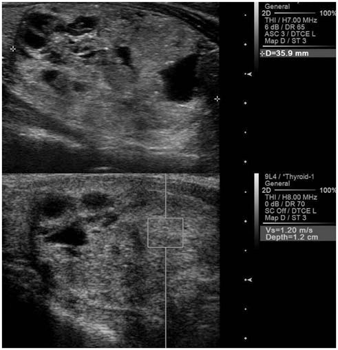
Fig. 2 Predominantly solid, isoechoic nodule shows low VTQ score (1.20 m/s) in ARFI evaluation. This nodule was confirmed as benign nodular hyperplasia. ARFI = acoustic radiation force impulse, VTQ = Virtual Touch tissue quantification
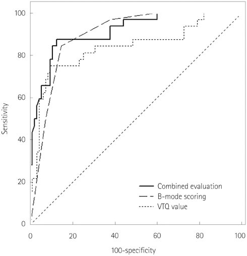
Fig. 3 Receiver operating characteristic curves for B-mode evaluation, VTQ value and combined evaluation. Area under the curve for combined evaluation (0.912) is higher than that for B-mode (0.895) or VTQ value (0.837). VTQ = Virtual Touch tissue quantification
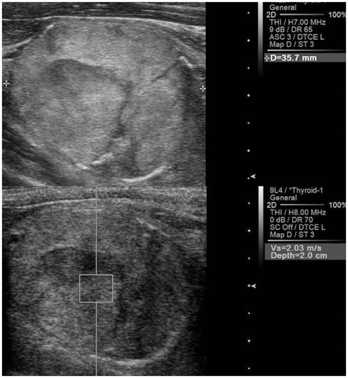
Fig. 4 Solid, heterogeneous isoechoic nodule shows relatively low VTQ value (2.03 m/s). This nodule was confirmed as follicular carcinoma. VTQ = Virtual Touch tissue quantification
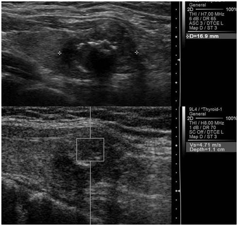
Fig. 5 Hypoechoic solid nodule with macrocalcification shows high VTQ value (4.71 m/s) in ARFI evaluation. This nodule was confirmed as benign nodule with calcific degeneration. ARFI = acoustic radiation force impulse, VTQ = Virtual Touch tissue quantification
Reference
-
1. Itoh A, Ueno E, Tohno E, Kamma H, Takahashi H, Shiina T, et al. Breast disease: clinical application of US elastography for diagnosis. Radiology. 2006; 239:341–350.2. Dighe M, Bae U, Richardson ML, Dubinsky TJ, Minoshima S, Kim Y. Differential diagnosis of thyroid nodules with US elastography using carotid artery pulsation. Radiology. 2008; 248:662–669.3. Hong Y, Liu X, Li Z, Zhang X, Chen M, Luo Z. Real-time ultrasound elastography in the differential diagnosis of benign and malignant thyroid nodules. J Ultrasound Med. 2009; 28:861–867.4. Rago T, Santini F, Scutari M, Pinchera A, Vitti P. Elastography: new developments in ultrasound for predicting malignancy in thyroid nodules. J Clin Endocrinol Metab. 2007; 92:2917–2922.5. Moon HJ, Sung JM, Kim EK, Yoon JH, Youk JH, Kwak JY. Diagnostic performance of gray-scale US and elastography in solid thyroid nodules. Radiology. 2012; 262:1002–1013.6. Kwak JY, Kim EK. Ultrasound elastography for thyroid nodules: recent advances. Ultrasonography. 2014; 33:75–82.7. Sun J, Cai J, Wang X. Real-time ultrasound elastography for differentiation of benign and malignant thyroid nodules: a meta-analysis. J Ultrasound Med. 2014; 33:495–502.8. Bojunga J, Herrmann E, Meyer G, Weber S, Zeuzem S, Friedrich-Rust M. Real-time elastography for the differentiation of benign and malignant thyroid nodules: a meta-analysis. Thyroid. 2010; 20:1145–1150.9. Zhai L, Palmeri ML, Bouchard RR, Nightingale RW, Nightingale KR. An integrated indenter-ARFI imaging system for tissue stiffness quantification. Ultrason Imaging. 2008; 30:95–111.10. Bojunga J, Dauth N, Berner C, Meyer G, Holzer K, Voelkl L, et al. Acoustic radiation force impulse imaging for differentiation of thyroid nodules. PLoS One. 2012; 7:e42735.11. Friedrich-Rust M, Romenski O, Meyer G, Dauth N, Holzer K, Grünwald F, et al. Acoustic Radiation Force Impulse-Imaging for the evaluation of the thyroid gland: a limited patient feasibility study. Ultrasonics. 2012; 52:69–74.12. Gu J, Du L, Bai M, Chen H, Jia X, Zhao J, et al. Preliminary study on the diagnostic value of acoustic radiation force impulse technology for differentiating between benign and malignant thyroid nodules. J Ultrasound Med. 2012; 31:763–771.13. Zhang YF, Xu HX, He Y, Liu C, Guo LH, Liu LN, et al. Virtual touch tissue quantification of acoustic radiation force impulse: a new ultrasound elastic imaging in the diagnosis of thyroid nodules. PLoS One. 2012; 7:e49094.14. Nightingale K, Soo MS, Nightingale R, Trahey G. Acoustic radiation force impulse imaging: in vivo demonstration of clinical feasibility. Ultrasound Med Biol. 2002; 28:227–235.15. Palmeri ML, Wang MH, Dahl JJ, Frinkley KD, Nightingale KR. Quantifying hepatic shear modulus in vivo using acoustic radiation force. Ultrasound Med Biol. 2008; 34:546–558.16. Zhan J, Diao XH, Chai QL, Chen Y. Comparative study of acoustic radiation force impulse imaging with real-time elastography in differential diagnosis of thyroid nodules. Ultrasound Med Biol. 2013; 39:2217–2225.17. Zhang FJ, Han RL. The value of acoustic radiation force impulse (ARFI) in the differential diagnosis of thyroid nodules. Eur J Radiol. 2013; 82:e686–e690.18. Kim HG, Moon HJ, Kwak JY, Kim EK. Diagnostic accuracy of the ultrasonographic features for subcentimeter thyroid nodules suggested by the revised American Thyroid Association guidelines. Thyroid. 2013; 23:1583–1589.19. Moon HJ, Kwak JY, Kim MJ, Son EJ, Kim EK. Can vascularity at power Doppler US help predict thyroid malignancy. Radiology. 2010; 255:260–269.20. Moon WJ, Baek JH, Jung SL, Kim DW, Kim EK, Kim JY, et al. Ultrasonography and the ultrasound-based management of thyroid nodules: consensus statement and recommendations. Korean J Radiol. 2011; 12:1–14.21. American Thyroid Association (ATA) Guidelines Taskforce on Thyroid Nodules and Differentiated Thyroid Cancer. Cooper DS, Doherty GM, Haugen BR, Kloos RT, Lee SL. Revised American Thyroid Association management guidelines for patients with thyroid nodules and differentiated thyroid cancer. Thyroid. 2009; 19:1167–1214.22. Asteria C, Giovanardi A, Pizzocaro A, Cozzaglio L, MorabitoA , Somalvico F, et al. US-elastography in the differential diagnosis of benign and malignant thyroid nodules. Thyroid. 2008; 18:523–531.23. Lyshchik A, Higashi T, Asato R, Tanaka S, Ito J, Mai JJ, et al. Thyroid gland tumor diagnosis at US elastography. Radiology. 2005; 237:202–211.24. Park SH, Kim SJ, Kim EK, Kim MJ, Son EJ, Kwak JY. Interobserver agreement in assessing the sonographic and elastographic features of malignant thyroid nodules. AJR Am J Roentgenol. 2009; 193:W416–W423.25. Nightingale K, Bentley R, Trahey G. Observations of tissue response to acoustic radiation force: opportunities for imaging. Ultrason Imaging. 2002; 24:129–138.26. Hou XJ, Sun AX, Zhou XL, Ji Q, Wang HB, Wei H, et al. The application of Virtual Touch tissue quantification (VTQ) in diagnosis of thyroid lesions: a preliminary study. Eur J Radiol. 2013; 82:797–801.
- Full Text Links
-
- Actions
-
Cited
- CITED
-
- Close
- Share
-
- Similar articles
-
- Ultrasound elastography for thyroid nodules: recent advances
- Diagnosis of Thyroid Nodules by Elastography
- Virtual Touch Tissue Quantification in the Differential Diagnosis of Benign and Malignant Thyroid Nodules
- Diagnostic Performance of a Combination of Shear Wave Elastography and B-Mode Ultrasonography in Differentiating Benign From Malignant Thyroid Nodules
- Advances in ultrasound elasticity imaging
