Pancreatic Arteriovenous Malformation as an Unusual Cause of Chronic Gastrointestinal Bleeding in a Patient with Early Gastric Cancer: Multimodality Imaging Spectrum with Pathologic Correlation
- Affiliations
-
- 1Department of Radiology, Chungnam National University Hospital, Chungnam National University School of Medicine, Daejeon, Korea. leeje290@gmail.com
- 2Department of Surgery, Chungnam National University Hospital, Chungnam National University School of Medicine, Daejeon, Korea.
- 3Department of Pathology, Chungnam National University Hospital, Chungnam National University School of Medicine, Daejeon, Korea.
- KMID: 2151772
- DOI: http://doi.org/10.13104/imri.2015.19.4.241
Abstract
- Arteriovenous malformation (AVM) of the pancreas is extremely rare, although it may be increasingly diagnosed due to the widespread use of cross-sectional imaging of the abdomen. Early diagnosis of this disease is important to prevent delay of treatment and resulting fatal complications. We report a rare case of pancreatic AVM in a 48-year-old man who presented with severe chronic anemia and early gastric cancer, which made diagnosis challenging. Imaging findings, including ultrasound, computed tomography, and magnetic resonance imaging, are shown, as well as the pathologic features.
Keyword
MeSH Terms
Figure
-

Fig. 1 Pancreatic arteriovenous malformation in a 41-year-old man, CT findings. (a) Axial scan, during the arterial phase, shows an irregularly tangled hypervascular lesion in the pancreatic head. (b) In the delayed phase, the lesion is difficult to define because of isodense enhancement relative to normal pancreatic parenchyma.

Fig. 2 Pancreatic arteriovenous malformation in a 41-year-old man, MRI findings. (a, b) T2-weighted imaging shows the characteristic clustered tubular signal void (arrows). (c-e) Axial scan during the arterial phase of dynamic T1-weighted imaging shows an irregularly tangled hypervascular lesion in the pancreatic head (c) and early enhancement of the dilated portal vein (d) and pancreaticoduodenal vein (arrow) that drained into the dilated portal vein (e).
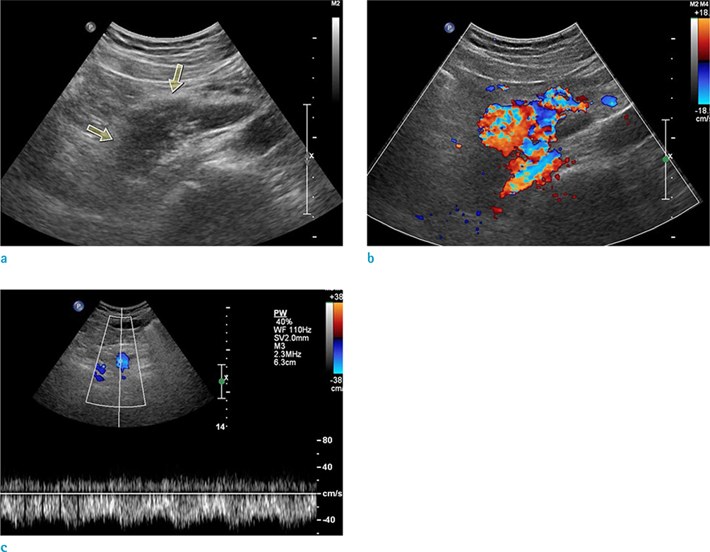
Fig. 3 Pancreatic arteriovenous malformation in a 41-year-old man, US findings. (a) A gray-scale ultrasonogram demonstrates an ill-marginated anechoic lesion (arrows) around the head of the pancreas. (b) On color Doppler ultrasonogram shows mosaic color flow pattern with large amount of color signals around the pancreas. (c) Spectral Doppler ultrasonogram of the main portal vein reveals pulsatile pattern, suggesting the draining role of the portal vein.

Fig. 4 Pancreatic arteriovenous malformation in a 41-year-old man, gross and microscopic findings. (a) A cut section of the pancreas shows multiple honeycomb-like dilated spaces. (b, c) Histopathologic examination of the resected pancreas reveals irregularly dilated angiodysplastic vessels in the pancreas (× 40, H&E staining) (b) and thick-walled artery and thin-walled vein connections with irregular duplication (arrows) (× 100, elastin staining) (c).
Reference
-
1. Rockey DC. Occult gastrointestinal bleeding. N Engl J Med. 1999; 341:38–46.2. Hansen W, Maximin S, Shriki JE, Bhargava P. Multimodality imaging of pancreatic arteriovenous malformation. Curr Probl Diagn Radiol. 2015; 44:105–109.3. Nishiyama R, Kawanishi Y, Mitsuhashi H, et al. Management of pancreatic arteriovenous malformation. J Hepatobiliary Pancreat Surg. 2000; 7:438–442.4. Meyer CT, Troncale FJ, Galloway S, Sheahan DG. Arteriovenous malformations of the bowel: an analysis of 22 cases and a review of the literature. Medicine (Baltimore). 1981; 60:36–48.5. Aida K, Nakamura H, Kihara Y, Abe S, Okamoto K, Otsuki M. Duodenal ulcer and pancreatitis associated with pancreatic arteriovenous malformation. Eur J Gastroenterol Hepatol. 2002; 14:551–554.6. Koito K, Namieno T, Nagakawa T, et al. Congenital arteriovenous malformation of the pancreas: its diagnostic features on images. Pancreas. 2001; 22:267–273.7. Makhoul F, Kaur P, Johnston TD, Jeon H, Gedaly R, Ranjan D. Arteriovenous malformation of the pancreas: a case report and review of literature. Int J Angiol. 2008; 17:211–213.8. Yoon JH, Han SS, Cha SS, Lee SJ. Color Doppler ultrasonography of a pancreatic arteriovenous malformation. J Ultrasound Med. 2005; 24:113–117.9. Walter JF, Chuang VP, Bookstein JJ, Reuter SR, Cho KJ, Pulmano CM. Angiography of massive hemorrhage secondary to pancreatic diseases. Radiology. 1977; 124:337–342.10. Chang S, Lim HK, Lee WJ, Choi D, Jang KT. Arteriovenous malformation of the pancreas in a patient with gastrointestinal bleeding: helical CT findings. Abdom Imaging. 2004; 29:259–262.
- Full Text Links
-
- Actions
-
Cited
- CITED
-
- Close
- Share
-
- Similar articles
-
- A Case of Pancreatic Arteriovenous Malformation with Portal Hypertension: Treatment with Transjugular Intrahepatic Portosystemic Shunt
- A Case of Arteriovenous Malformation of the Pancreas
- A Case of Duodenal Ulcer Bleeding caused by Pancreatic Arteriovenous Malformation
- A Case of a Spontaneous Hemoperitoneum as the Presentation of a Gastric Arteriovenous Malformation
- A case of hemosuccus pancreaticus in arteriovenous malformation
