Atypical Intramuscular Myxoma of the Lumbosacral Paraspinal Muscle: The First Case Report in Asian
- Affiliations
-
- 1Department of Neurosurgery, College of Medicine, Incheon St. Mary's Hospital, The Catholic University of Korea, Incheon, Korea. rhother@hanmail.net
- 2Department of Pathology, College of Medicine, Incheon St. Mary's Hospital, The Catholic University of Korea, Incheon, Korea.
- KMID: 2151163
- DOI: http://doi.org/10.3340/jkns.2015.58.6.566
Abstract
- Intramuscular myxoma (IM) is a benign neoplasm of mesenchymal origin. We report a rare case of IM which was located in the lumbosacral paraspinal muscles. A 62-year-old female patient presented with progressive low back pain for 2 months, and the radiologic findings showed a large mass (4.0x3.5x6.5 cm) in the right lumbosacral paraspinal area. Total resection of the tumor was performed and the symptom was nearly resolved after surgery. Although the immuno-histopathological analysis was consistent with IM, there were some different findings from typical pathological characteristics of IM in this case. Firstly, the symptomatic change of the mass took relatively short time (less than 3 months), and this change was accompanied by partial calcification inside the mass. Moreover, iatrogenic interruption of paravertebral muscle by the other previous operation might be the promoting factor of the fibrous dysplasia, which can explain the pathogenesis of IM. To our knowledge, this is the eighth case of the lumbar paraspinal myxoma reported in the literatures and the first case in Asian population.
Keyword
MeSH Terms
Figure
-
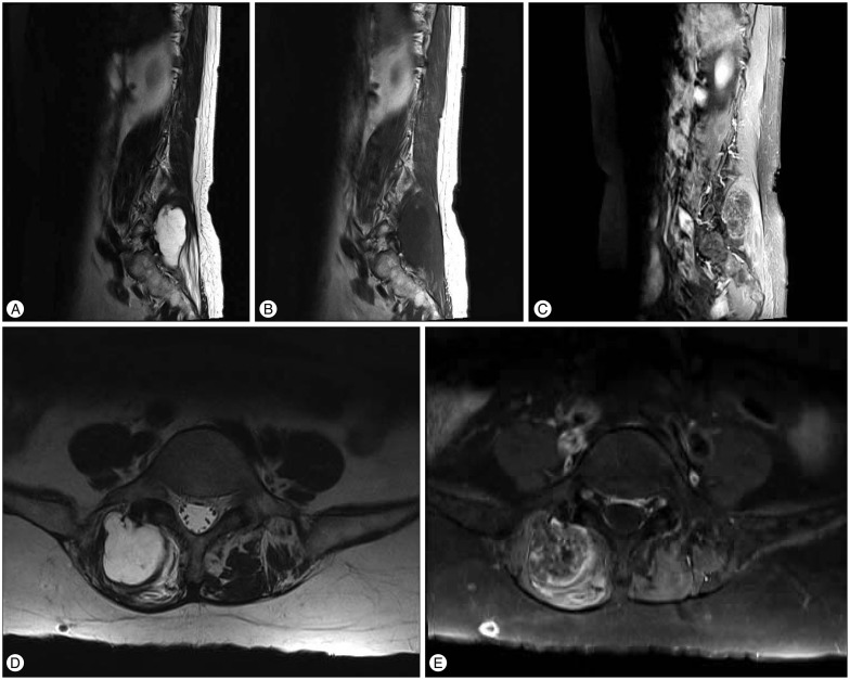
Fig. 1 Preoperative magnetic resonance images. The multi-lobulated mass was located within the right lumbosacral paraspinal muscles in contact with the posterior elements of the lumbosacral vertebra. This large mass measured approximately 4.0×3.5×6.5 cm in size, and the lesion was hypointense on T1-weighted images (B) and hyperintense on T2-weighted images (A and D). On the post-contrast images (C and E), there was heterogenously enhancing foci inside the mass with peripheral and septal enhancement.
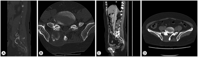
Fig. 2 Comparing the preoperative lumbar spine CT (A and B) with the previous abdominal CT (C and D). Note that the calcification in the inferior portion of the mass (A and B), which was not detected on the abdominal CT examined 4 months ago.
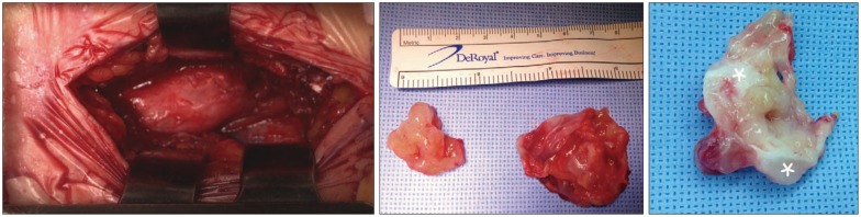
Fig. 3 Intraoperative photographs. The mass was identified through a paramedian longitudinal incision and was excised en bloc. Partial calcification was detected inside the tumor (asterisks).
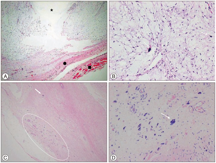
Fig. 4 Microscopic findings of the paraspinal myxomas. A : Myxoid hypocelluar lesion with fibrous capsule (circle). Note the microcystic change in mass (star). Degenerated skeletal muscle fiber (square) also identified (H&E, ×40). B : Bland-looking spindle to stellate cells in bluish myxoid stroma. Note the pyknotic nuclei and microcalcification (arrow) (H&E, ×200). C : Numerous karyorrhectic nuclei (circle) and microcalcification (arrow) in periphery-capsular area of tumor, which was seen in CT (H&E, ×40). D : Numerous karyorrhectic nuclei and microcalcification (arrow) in periphery-capsular area of tumor (H&E, ×200).
Reference
-
1. Bancroft LW, Kransdorf MJ, Menke DM, O'Connor MI, Foster WC. Intramuscular myxoma : characteristic MR imaging features. AJR Am J Roentgenol. 2002; 178:1255–1259. PMID: 11959742.2. Canalis RF, Smith GA, Konrad HR. Myxomas of the head and neck. Arch Otolaryngol. 1976; 102:300–305. PMID: 1267724.
Article3. Crankson SJ, Al Namshan M, Al Mane K, Bamefleh H. Intramuscular myxoma : a rare neck mass in a child. Pediatr Radiol. 2002; 32:120–122. PMID: 11819080.4. Doyle LA, Möller E, Dal Cin P, Fletcher CD, Mertens F, Hornick JL. MUC4 is a highly sensitive and specific marker for low-grade fibromyxoid sarcoma. Am J Surg Pathol. 2011; 35:733–741. PMID: 21415703.
Article5. Enzinger FM. Intramuscular myxoma; a review and follow-up study of 34 cases. Am J Clin Pathol. 1965; 43:104–113. PMID: 14253111.
Article6. Falavigna A, Righesso O, Volquind D, Teles AR. Intramuscular myxoma of the cervical paraspinal muscle. Eur Spine J. 2009; 18(Suppl 2):245–249. PMID: 19301043.
Article7. Feldman PS. A comparative study including ultrastructure of intramuscular myxoma and myxoid liposarcoma. Cancer. 1979; 43:512–525. PMID: 421179.
Article8. Guppy KH, Wagner F, Tawk R, Gallagher L. Intramuscular myxoma causing lumbar radiculopathy. Case report and review of the literature. J Neurosurg. 2001; 95(2 Suppl):260–263. PMID: 11599850.9. Kamoun N, Zouari M, Siala M, Karray S, Douik M, Litaiem T, et al. [Intramuscular myxoma. Apropos of two cases]. Rev Chir Orthop Reparatrice Appar Mot. 1997; 83:278–282. PMID: 9255366.10. Kindblom LG, Stener B, Angervall L. Intramuscular myxoma. Cancer. 1974; 34:1737–1744. PMID: 4426032.
Article11. Liguoro D, Viejo-Fuertes D, Vital A, San Galli F, Dautheribes M, Guerin J. [Intramuscular myxoma. A case of myxoma of the spinal erector muscle]. Neurochirurgie. 1999; 45:54–57. PMID: 10374236.12. Manoharan SR, Shaw AB, Arnold CA, Farhadi HF. Infiltrative intramuscular myxoma of the cervical spine : a case report. Spine J. 2015; 15:e1–e4. PMID: 25264316.13. Nielsen GP, O'Connell JX, Rosenberg AE. Intramuscular myxoma : a clinicopathologic study of 51 cases with emphasis on hypercellular and hypervascular variants. Am J Surg Pathol. 1998; 22:1222–1227. PMID: 9777984.14. Ohla V, Ciarlini PD, Goldsmith JD, Kasper EM. Cellular myxoma of the lumbar spine. Surg Neurol Int. 2013; 4:82. PMID: 23869282.
Article15. Rashid A, Abdul-Jabar HB, Karmani S, Rezajooi K, Casey AT. Giant paravertebral myxoma. Eur Spine J. 2011; 20(Suppl 2):S138–S142. PMID: 20495934.
Article16. Shugar JM, Som PM, Meyers RJ, Schaeffer BT. Intramuscular head and neck myxoma : report of a case and review of the literature. Laryngoscope. 1987; 97:105–107. PMID: 3796168.17. Stinchcombe S, Kochhar R, Malkan D. Intramuscular myxoma of the paraspinal musculature. J Med Cases. 2010; 1:42–46.
Article18. Stout AP. Myxoma, the tumor of primitive mesenchyme. Ann Surg. 1948; 127:706–719. PMID: 18917127.19. Taggarshe D, Raheja S, Yoo S, Mittal V. Intramuscular myxoma : a rare back mass. Am Surg. 2010; 76:1303–1304. PMID: 21140704.20. Tahmouresie A, Farmer PM, Stokes N. Paraspinal myxoma with spinal cord compression. Case report. J Neurosurg. 1981; 54:542–544. PMID: 7205357.21. Tataryn Z, Tracy J, Tsang C, Wu J, Heilman CB, Wein RO. Intramuscular myxoma of the cervical paraspinal musculature : case report and review of the literature. Am J Otolaryngol. 2015; 36:273–276. PMID: 25481300.
Article22. Tomich CE. Oral focal mucinosis. A clinicopathologic and histochemical study of eight cases. Oral Surg Oral Med Oral Pathol. 1974; 38:714–724. PMID: 4140487.
