Bilateral Bone Marrow Edema Syndrome of the Femoral Head with a Unique Onset: A Case Report
- Affiliations
-
- 1Department of Orthopedic Surgery, National Police Hospital, Seoul, Korea. segaba1@naver.com
- KMID: 2150516
- DOI: http://doi.org/10.5371/hp.2015.27.4.273
Abstract
- Bone marrow edema syndrome (BMES) is a rare condition which mainly affects the hip area. The etiology and pathogenesis of BMES is still unclear. Pain near the affected area, regional osteoporosis, bone marrow edema (identified using magnetic resonance imaging) and spontaneous regression within 6-12 months are the main characteristics of BMES. In this case, a 52-year-old male was diagnosed with BMES of the right hip followed by spontaneous subsiding of symptoms. After 3 years, and under nearly the same social and physical conditions, he was admitted again with newly developed left hip pain and again diagnosed with BMES. We report this rare case since a similar one has not been previously reported in the domestic literature and may be considered valuable for basic research relating to the pathogenesis of BMES.
Keyword
Figure
-

Fig. 1 Imaging studies of a 52-year-old male at the first clinical visit for right hip pain. (A) Plain radiograph shows no significant abnormality around the hips. (B) Bone scan shows marked, homogenous hot uptake centered in the femoral head. (C) Magnetic resonance imaging shows low to intermediate signal intensity in the right femoral head, neck, greater and lesser trochanteric area in T1-weighted images, and high signal intensity in the same areas in T2-weighted images. About 1.5 cm-long fluid collection in the insertion of the iliopsoas muscle is observed at the anterior area of the right femur neck.
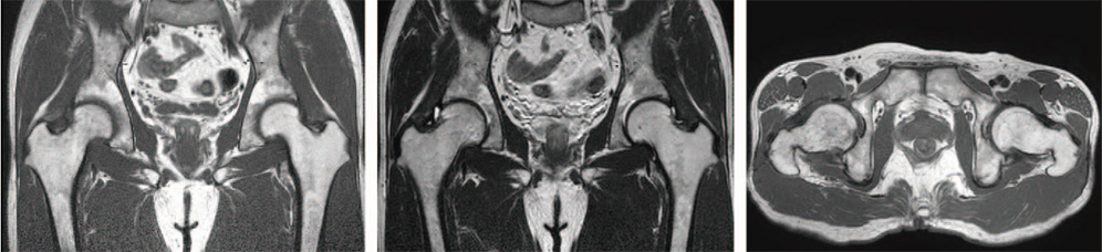
Fig. 2 Follow-up magnetic resonance imaging obtained after 3 months shows no definitive abnormal finding.
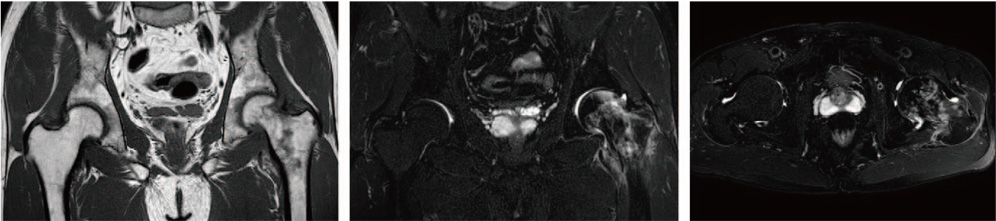
Fig. 3 Imaging studies of the same patient at the second clinical visit for the left hip pain after 3 years. Magnetic resonance imaging (MRI) of the hips shows low to intermediate signal intensity in the left femoral head, neck, greater and lesser trochanteric area in T1-weighted images and high signal intensity in the same areas in T2-weighted images. MRI patterns are similar with previous MRI of the right hip.
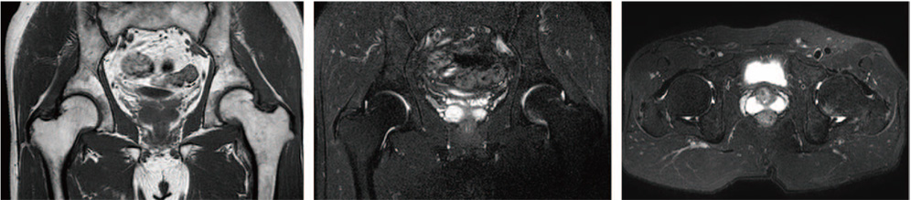
Fig. 4 Follow-up magnetic resonance imaging obtained after 2 months demonstrates significant improvement compared with Fig. 3.
Reference
-
1. Curtiss PH Jr, Kincaid WE. Transitory demineralization of the hip in pregnancy. A report of three cases. J Bone Joint Surg Am. 1959; 41-A:1327–1333.2. Solomon L. Bone-marrow oedema syndrome. J Bone Joint Surg Br. 1993; 75:175–176.
Article3. Hayes CW, Conway WF, Daniel WW. MR imaging of bone marrow edema pattern: transient osteoporosis, transient bone marrow edema syndrome, or osteonecrosis. Radiographics. 1993; 13:1001–1011.
Article4. Hofmann S, Engel A, Neuhold A, Leder K, Kramer J, Plenk H Jr. Bone-marrow oedema syndrome and transient osteoporosis of the hip. An MRI-controlled study of treatment by core decompression. J Bone Joint Surg Br. 1993; 75:210–216.
Article5. Resnick D, Niwayama G. Transient osteoporosis of the hip. In : Resnick D, Niwayama G, editors. Diagnosis of bone and joint disorders. 2nd ed. Philadelphia: Saunders;1988. p. 2013–2016.6. Gao F, Sun W, Li Z, Guo W, Kush N, Ozaki K. Intractable bone marrow edema syndrome of the hip. Orthopedics. 2015; 38:e263–e270.
Article7. Patel S. Primary bone marrow oedema syndromes. Rheumatology (Oxford). 2014; 53:785–792.
Article8. Niimi R, Sudo A, Hasegawa M, Fukuda A, Uchida A. Changes in bone mineral density in transient osteoporosis of the hip. J Bone Joint Surg Br. 2006; 88:1438–1440.
Article
- Full Text Links
-
- Actions
-
Cited
- CITED
-
- Close
- Share
-
- Similar articles
-
- Bone Marrow Pressure of the Femoral Heads of Korean Adults
- Bone Marrow Edema Syndrome of the Foot and Ankle in Adolescents
- The Early diagnostic Significance of Bone Marrow Pressure in Osteonecrosis of the Femoral Head
- Occurrence of Marrow Edema in Early Stage Osteonecrosis of the Femoral Head: a Prospective Study with Repeated MR Imagings
- Bone Marrow Pressure Study in Ostoenecrosis of the Femoral Head
