Single Balloon Enteroscopy-Assisted Endoscopic Retrograde Cholangiopancreatography in Patients Who Underwent a Gastrectomy with Roux-en-Y Anastomosis: Six Cases from a Single Center
- Affiliations
-
- 1Department of Gastroenterology, Asan Digestive Disease Research Institute, Asan Medical Center, University of Ulsan College of Medicine, Seoul, Korea. dhyang@amc.seoul.kr
- KMID: 2148561
- DOI: http://doi.org/10.5946/ce.2015.48.5.452
Abstract
- Patients with altered anatomy such as a Roux-en-Y anastomosis often present with various pancreaticobiliary problems requiring therapeutic intervention. However, a conventional endoscopic approach to the papilla is very difficult owing to the long afferent limb and acute angle of a Roux-en-Y anastomosis. Balloon-assisted enteroscopy can be used for endoscopic retrograde cholangiopancreatography (ERCP) in patients with altered anatomy. We experienced six cases of Roux-en-Y anastomosis with biliary problems, and attempted ERCP using single balloon enteroscopy (SBE). SBE insertion followed by replacement with a conventional endoscope was attempted in five of six patients. The papilla was successfully approached using SBE in all cases. However, therapeutic intervention was completed in only three cases because of poor maneuverability caused by postoperative adhesion. We conclude that in patients with Roux-en-Y anastomosis, the ampulla can be readily accessed with SBE, but longer dedicated accessories are necessary to improve this therapeutic intervention.
Keyword
MeSH Terms
Figure
-
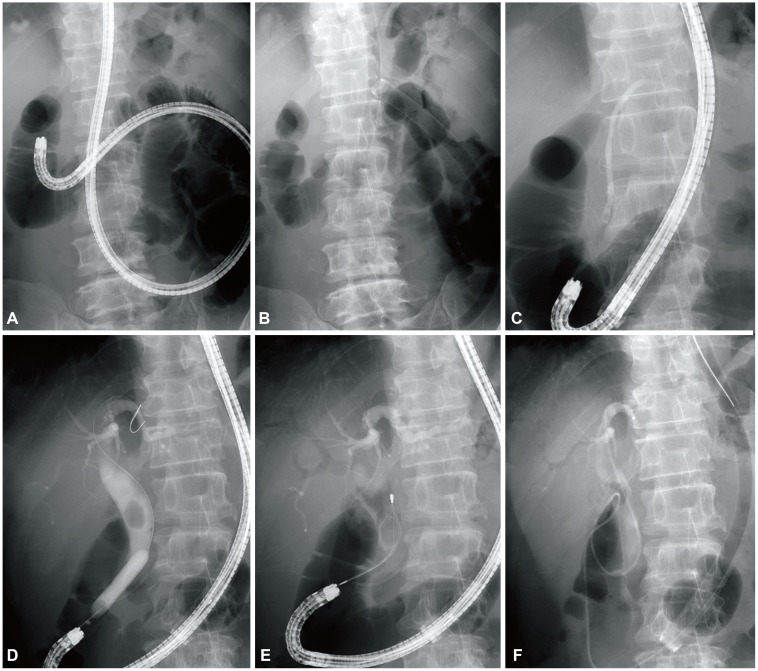
Fig. 1 A 58-year-old man who received a total gastrectomy with a Roux-en-Y anastomosis had a 17-mm brown stone removed by single balloon enteroscope (SBE)-assisted endoscopic retrograde cholangiopancreatography (ERCP). (A) Simple radiography showing the tip of the SBE reaching the end of the afferent loop. (B) The SBE was withdrawn leaving the overtube. (C) An esophagogastroduodenoscope was inserted successfully through the in-dwelling overtube. (D) A cholangiogram showing a diffuse dilatation and a 17-mm filling defect in the common bile duct. Endoscopic papillary dilatation was performed with the controlled radial expansion balloon. (E) The stone was removed using a mechanical lithotripter and a retrieval balloon. (F) An endoscopic nasobiliary drain was inserted and ERCP was completed successfully.
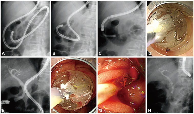
Fig. 2 A 78-year-old man who received a prior total gastrectomy with a Roux-en-Y anastomosis had small brown stones successfully removed by single balloon enteroscope (SBE)-assisted endoscopic retrograde cholangiopancreatography (ERCP). (A) The SBE reached the afferent loop. (B) The enteroscope was 80 cm in length. (C) While inserting the esophagogastroduodenoscope, an acute angulation was made 25 cm distal to the overtube. After the collapsed site was dilated by the controlled radial expansion (CRE) balloon, a conventional endoscope could be inserted into the overtube. (D) Endoscopic view; the CRE balloon dilated the acute angulation. (E) Dilatation of the common bile duct was noted on the cholangiogram. (F) Endoscopic papillary dilatation was performed with a CRE balloon. (G) Small brown stones were removed using the retrieval balloon. (H) An endoscopic nasobiliary drainage was inserted and ERCP was completed successfully.
Cited by 1 articles
-
Direct Insertion of a Short-Type Single-Balloon Enteroscope and Using a Stent Retriever to Treat Difficult Bile Duct Stones in Surgically Altered Anatomy
Takashi Sasaki, Naoki Sasahira
Clin Endosc. 2021;54(6):937-938. doi: 10.5946/ce.2020.145.
Reference
-
1. Koornstra JJ, Fry L, Mönkemüller K. ERCP with the balloon-assisted enteroscopy technique: a systematic review. Dig Dis. 2008; 26:324–329. PMID: 19188723.
Article2. Moreels TG. Altered anatomy: enteroscopy and ERCP procedure. Best Pract Res Clin Gastroenterol. 2012; 26:347–357. PMID: 22704576.
Article3. Itoi T, Ishii K, Sofuni A, et al. Single-balloon enteroscopy-assisted ERCP in patients with Billroth II gastrectomy or Roux-en-Y anastomosis (with video). Am J Gastroenterol. 2010; 105:93–99. PMID: 19809409.
Article4. Hintze RE, Adler A, Veltzke W, Abou-Rebyeh H. Endoscopic access to the papilla of Vater for endoscopic retrograde cholangiopancreatography in patients with billroth II or Roux-en-Y gastrojejunostomy. Endoscopy. 1997; 29:69–73. PMID: 9101141.
Article5. Elton E, Hanson BL, Qaseem T, Howell DA. Diagnostic and therapeutic ERCP using an enteroscope and a pediatric colonoscope in long-limb surgical bypass patients. Gastrointest Endosc. 1998; 47:62–67. PMID: 9468425.
Article6. Wright BE, Cass OW, Freeman ML. ERCP in patients with long-limb Roux-en-Y gastrojejunostomy and intact papilla. Gastrointest Endosc. 2002; 56:225–232. PMID: 12145601.
Article7. Emmett DS, Mallat DB. Double-balloon ERCP in patients who have undergone Roux-en-Y surgery: a case series. Gastrointest Endosc. 2007; 66:1038–1041. PMID: 17963892.
Article8. Mönkemüller K, Fry LC, Bellutti M, Neumann H, Malfertheiner P. ERCP with the double balloon enteroscope in patients with Roux-en-Y anastomosis. Surg Endosc. 2009; 23:1961–1967. PMID: 19067052.
Article9. Parlak E, Ciçek B, Dişibeyaz S, et al. Endoscopic retrograde cholangiography by double balloon enteroscopy in patients with Roux-en-Y hepaticojejunostomy. Surg Endosc. 2010; 24:466–470. PMID: 19585072.
Article10. Mönkemüller K, Fry LC, Bellutti M, Neumann H, Malfertheiner P. ERCP using single-balloon instead of double-balloon enteroscopy in patients with Roux-en-Y anastomosis. Endoscopy. 2008; 40(Suppl 2):E19–E20. PMID: 18278720.
Article11. Kianička B, Lata J, Novotný I, Dítě P, Vaníček J. Single balloon enteroscopy for endoscopic retrograde cholangiography in patients with Roux-en-Y hepaticojejuno anastomosis. World J Gastroenterol. 2013; 19:8047–8055. PMID: 24307799.12. Saleem A, Baron TH, Gostout CJ, et al. Endoscopic retrograde cholangiopancreatography using a single-balloon enteroscope in patients with altered Roux-en-Y anatomy. Endoscopy. 2010; 42:656–660. PMID: 20589594.
Article13. Tomizawa Y, Sullivan CT, Gelrud A. Single balloon enteroscopy (SBE) assisted therapeutic endoscopic retrograde cholangiopancreatography (ERCP) in patients with roux-en-y anastomosis. Dig Dis Sci. 2014; 59:465–470. PMID: 24185681.
Article14. Kawamura T, Mandai K, Uno K, Yasuda K. Does single-balloon enteroscopy contribute to successful endoscopic retrograde cholangiopancreatography in patients with surgically altered gastrointestinal anatomy? ISRN Gastroenterol. 2013; 2013:214958. PMID: 23762573.
Article15. Moreels TG, Pelckmans PA. Comparison between double-balloon and single-balloon enteroscopy in therapeutic ERC after Roux-en-Y entero-enteric anastomosis. World J Gastrointest Endosc. 2010; 2:314–317. PMID: 21160763.
Article16. Kawamura T, Uno K, Suzuki A, et al. Clinical usefulness of a short-type, prototype single-balloon enteroscope for endoscopic retrograde cholangiopancreatography in patients with altered gastrointestinal anatomy: preliminary experiences. Dig Endosc. 2015; 27:82–86. PMID: 25040667.
Article17. Siddiqui AA, Chaaya A, Shelton C, et al. Utility of the short double-balloon enteroscope to perform pancreaticobiliary interventions in patients with surgically altered anatomy in a US multicenter study. Dig Dis Sci. 2013; 58:858–864. PMID: 22975796.
Article
- Full Text Links
-
- Actions
-
Cited
- CITED
-
- Close
- Share
-
- Similar articles
-
- Endoscopic ultrasound-directed transgastric endoscopic retrograde cholangiopancreatography for patients with Roux-en-Y gastric bypass anatomy: technical overview
- Endoscopic Retrograde Cholangiopancreatography in Post Gastrectomy Patients
- Impact of Scope Exchange from a Long Single Balloon Enteroscope to a Gastroscope during Enteroscopy-Assisted Endoscopic Retrograde Cholangiopancreatography in Patients with Surgically Altered Anatomy
- Cap-assisted ERCP in Surgically Altered Anatomy
- Recent advances of endoscopic retrograde cholangiopancreatography in surgically altered anatomy
