Mobile Sensor Application for Kinematic Detection of the Knees
- Affiliations
-
- 1Biomedical Engineering Program, Faculty of Engineering, Chulalongkorn University, Bangkok, Thailand. toss.jay@gmail.com
- 2Department of Rehabilitation Medicine, Faculty of Medicine, Chulalongkorn University, Bangkok, Thailand.
- 3Department of Electrical Engineering, Faculty of Engineering, Chulalongkorn University, Bangkok, Thailand.
- KMID: 2148231
- DOI: http://doi.org/10.5535/arm.2015.39.4.599
Abstract
OBJECTIVE
To correctly measure the knee joint angle, this study utilized a Qualisys motion capture system and also used it as the reference to assess the validity of the study's Inertial Measurement Unit (IMU) system that consisted of four IMU sensors and the Knee Angle Recorder software. The validity was evaluated by the root mean square (RMS) of different angles and the intraclass correlation coefficient (ICC) values between the Qualisys system and the IMU system.
METHODS
Four functional knee movement tests for ten healthy participants were investigated, which were the knee flexion test, the hip and knee flexion test, the forward step test and the leg abduction test, and the walking test.
RESULTS
The outcomes of the knee flexion test, the hip and knee flexion test, the forward step test, and the walking test showed that the RMS of different angles were less than 6degrees. The ICC values were in the range of 0.84 to 0.99. However, the leg abduction test showed a poor correlation in the measurement of the knee abduction-adduction movement.
CONCLUSION
The IMU system used in this study is a new good method to measure the knee flexion-extension movement.
Keyword
Figure
-
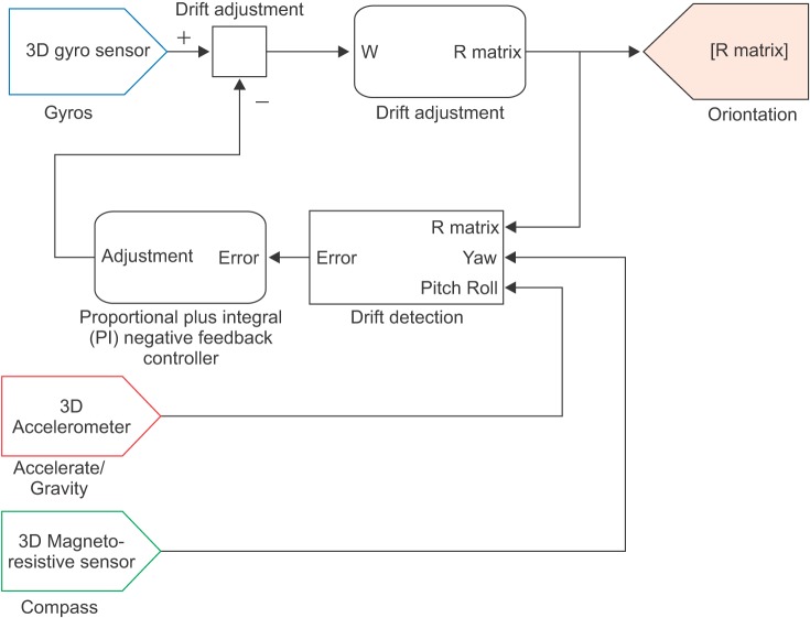
Fig. 1 A block diagram of the Direction Cosine Matrix process. Adapted from Premerlani W, Bizard P. Direction cosine matrix IMU: theory (http://gentlenav.googlecode.com/files/DCMDraft2.pdf), with permission [10].
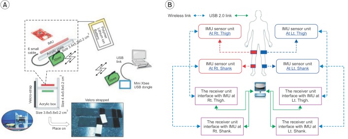
Fig. 2 (A) shows the Inertial Measurement Unit (IMU) devices developed for this study. (B) is the diagram of the IMU system used in this study.
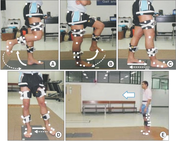
Fig. 3 The movement tests used in this study. (A) The knee flexion test, (B) the hip and knee flexion test, (C) the forward step test, (D) the leg abduction test, and (E) the walking test along a walkway.
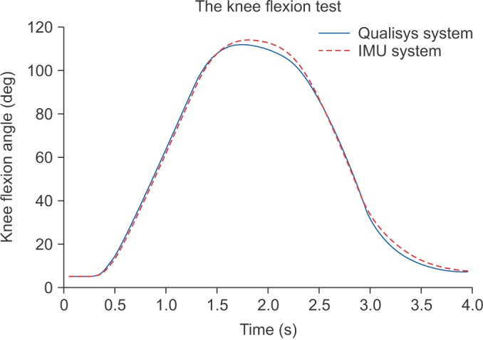
Fig. 4 The line graphs of knee flexion angles versus time during the knee flexion test, which is a comparison between the Qualisys system and the Inertial Measurement Unit (IMU) system (the data is from participant no. 1, the right leg).
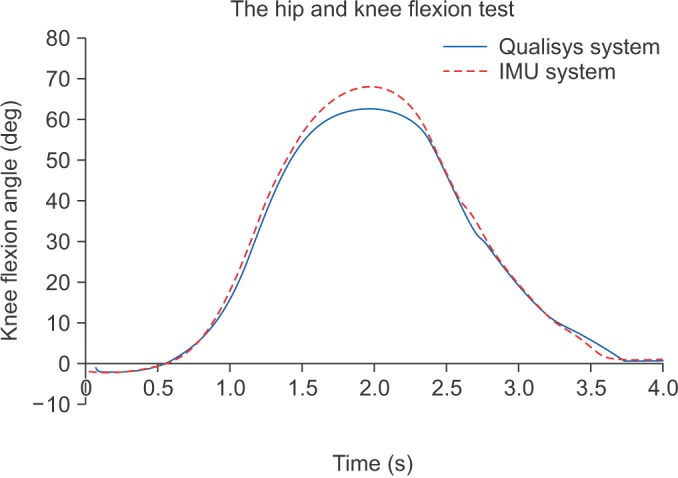
Fig. 5 The line graphs of knee flexion angles versus time during the hip and knee flexion test, which is a comparison between the Qualisys system and the Inertial Measurement Unit (IMU) system (the data is from participant no. 10, the right leg).
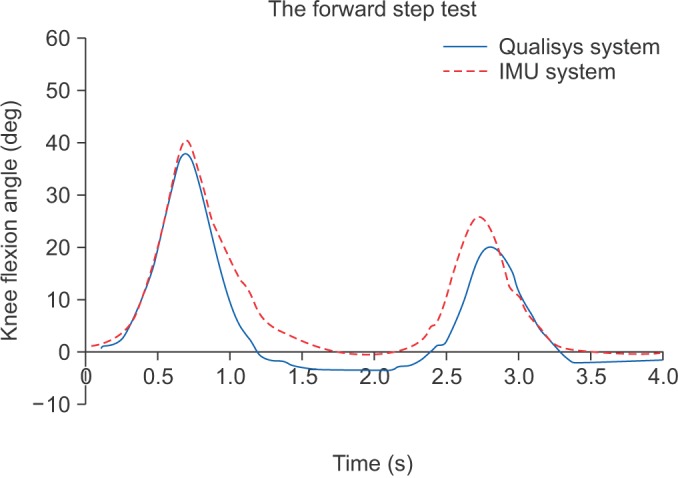
Fig. 6 The line graphs of knee flexion angles versus time during the forward step test, which is a comparison between the Qualisys system and Inertial Measurement Unit (IMU) system (the data is from the participant no. 3, the left leg).
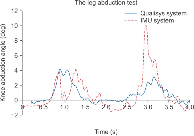
Fig. 7 The line graphs of knee abduction angles versus time during the leg abduction test, which is a comparison between the Qualisys system and Inertial Measurement Unit (IMU) system (the data is from the participant no. 9, the left leg).

Fig. 8 The line graphs of knee flexion angles versus time in a gait cycle during the walking test, which is a comparison between the Qualisys system and Inertial Measurement Unit (IMU) system (the data is from the participant no. 8, both legs).
Reference
-
1. Favre J, Aissaoui R, Jolles BM, de Guise JA, Aminian K. Functional calibration procedure for 3D knee joint angle description using inertial sensors. J Biomech. 2009; 42:2330–2335. PMID: 19665712.
Article2. Luinge HJ, Veltink PH. Measuring orientation of human body segments using miniature gyroscopes and accelerometers. Med Biol Eng Comput. 2005; 43:273–282.
Article3. Favre J, Jolles BM, Aissaoui R, Aminian K. Ambulatory measurement of 3D knee joint angle. J Biomech. 2008; 41:1029–1035. PMID: 18222459.
Article4. Mayagoitia RE, Nene AV, Veltink PH. Accelerometer and rate gyroscope measurement of kinematics: an inexpensive alternative to optical motion analysis systems. J Biomech. 2002; 35:537–542. PMID: 11934425.
Article5. Dejnabadi H, Jolles BM, Aminian K. A new approach to accurate measurement of uniaxial joint angles based on a combination of accelerometers and gyroscopes. IEEE Trans Biomed Eng. 2005; 52:1478–1484.
Article6. Cooper G, Sheret I, McMillan L, Siliverdis K, Sha N, Hodgins D, et al. Inertial sensor-based knee flexion/extension angle estimation. J Biomech. 2009; 42:2678–2685.
Article7. Takeda R, Tadano S, Natorigawa A, Todoh M, Yoshinari S. Gait posture estimation using wearable acceleration and gyro sensors. J Biomech. 2009; 42:2486–2494.
Article8. Takeda R, Tadano S, Todoh M, Morikawa M, Nakayasu M, Yoshinari S. Gait analysis using gravitational acceleration measured by wearable sensors. J Biomech. 2009; 42:223–233. PMID: 19121522.
Article9. Chardonnens J, Favre J, Cuendet F, Gremion G, Aminian K. A system to measure the kinematics during the entire ski jump sequence using inertial sensors. J Biomech. 2013; 46:56–62. PMID: 23123073.
Article10. Chardonnens J, Favre J, Le Callennec B, Cuendet F, Gremion G, Aminian K. Automatic measurement of key ski jumping phases and temporal events with a wearable system. J Sports Sci. 2012; 30:53–61. PMID: 22168430.
Article11. Rouhani H, Favre J, Crevoisier X, Aminian K. Ambulatory measurement of ankle kinetics for clinical applications. J Biomech. 2011; 44:2712–2718. PMID: 21851944.
Article12. Premerlani W, Bizard P. Direction cosine matrix IMU: theory [Internet]. place unknown: publisher unknown;2009. cited 2015 Jul 15. Available from: gentlenav.googlecode.com/files/DCMDraft2.pdf.13. Jaysrichai T, Suputtitada A, Khovidhungij W, Chanwimalueang T. Application of inertial measurement units for angular motion detection. In : Proceedings of the 7th International Convention on Rehabilitation Engineering and Assistive Technology; 2013 Aug 29-31; Seoul, Korea.14. Luinge HJ, Veltink PH. Inclination measurement of human movement using a 3-D accelerometer with autocalibration. IEEE Trans Neural Syst Rehabil Eng. 2004; 12:112–121. PMID: 15068194.
Article15. Morris JR. Accelerometry: a technique for the measurement of human body movements. J Biomech. 1973; 6:729–736. PMID: 4757490.16. Tong K, Granat MH. A practical gait analysis system using gyroscopes. Med Eng Phys. 1999; 21:87–94. PMID: 10426508.
Article17. Favre J, Luthi F, Jolles BM, Siegrist O, Najafi B, Aminian K. A new ambulatory system for comparative evaluation of the three-dimensional knee kinematics, applied to anterior cruciate ligament injuries. Knee Surg Sports Traumatol Arthrosc. 2006; 14:592–604. PMID: 16421753.
Article18. Aminian K, Najafi B. Capturing human motion using body-fixed sensors: outdoor measurement and clinical applications. Comput Animat Virtual Worlds. 2004; 15:79–94.
Article
- Full Text Links
-
- Actions
-
Cited
- CITED
-
- Close
- Share
-
- Similar articles
-
- The Effects of an Interactive Nursing Skills Mobile Application on Nursing Students' Knowledge, Self-efficacy, and Skills Performance: A Randomized Controlled Trial
- Development of a mobile application focusing on developmental support care for Korean infants born prematurely: a methodological study
- Measurement of Knee Rotation Angles Using a Smartphone Application: An Experimental Study of Porcine Knees
- Development and User Satisfaction of a Mobile Phone Application for Image-based Dietary Assessment
- Development and Validation of a Postpartum Care Mobile Application for First-time Mothers
