J Korean Surg Soc.
2010 Oct;79(4):306-309. 10.4174/jkss.2010.79.4.306.
Left Adrenal Sarcoidosis
- Affiliations
-
- 1Department of Surgery, St. Vincent's Hospital, The Catholic University of Korea, Suwon, Korea. hmcho@catholic.ac.kr
- 2Department of Pathology, St. Vincent's Hospital, The Catholic University of Korea, Suwon, Korea.
- 3Department of Radiology, St. Vincent's Hospital, The Catholic University of Korea, Suwon, Korea.
- KMID: 2145034
- DOI: http://doi.org/10.4174/jkss.2010.79.4.306
Abstract
- We present a 50-year-old woman with left adrenal sarcoidosis. She visited our hospital for right upper quadrant discomfort; she was then evaluated for right upper quadrant discomfort. She had no abnormal findings in the laboratory examination, including hormone study, but a mass was detected at left adrenal gland, incidentally. Initially, we thought the mass as nonfunction adrenal adenoma. After she had undergone laparoscopic left adrenalectomy, she was diagnosed with left adrenal sarcoidosis from her histological findings. Adrenal sarcoidosis is a very rare disease. This case provides insight to the experience of left adrenal sarcoidosis.
Keyword
Figure
-
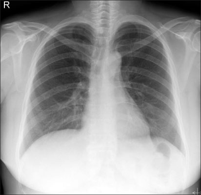
Fig. 1 Chest X-ray. There is no abnormal finding.

Fig. 2 Abdominal CT. There is multi-nodular thickening of left adrenal gland (less than 2×1.4 cm) suggesting adrenal adenoma or malignant lesion.

Fig. 3 PET CT. Images showed a 2×1.4 cm sized mass with increased FDG uptake (mSUV of 7.9) at left adrenal gland, suggesting malignant tumor at left adrenal gland.
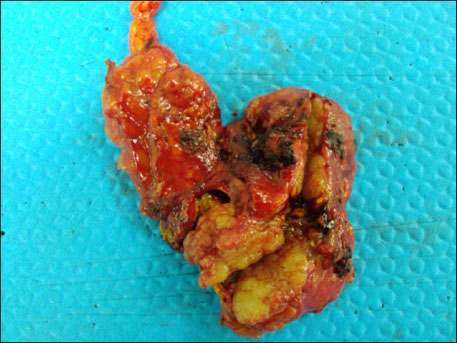
Fig. 4 The specimen consists of a portion of tan-colored tissue, measuring 5.5×3.2×2.2 cm.

Fig. 5 There are noncaseating granulomas mainly composed of epithelioid cell, with scattered Langhans' giant cells and lymphocytes. Necrosis is absent and no organisms were identified. At the periphery of the lesion, the adrenal cortex is seen (H&E, ×200).
Reference
-
1. Porter N, Beynon HL, Randeva HS. Endocrine and reproductive manifestations of sarcoidosis. QJM. 2003. 96:553–561.2. Judson MA. Sarcoidosis: clinical presentation, diagnosis, and approach to treatment. Am J Med Sci. 2008. 335:26–33.3. Gostiljac DM, Dordević PB, Maric-zivković J, Canović F. Sarcoidosis localized in endocrine glands. Med Pregl. 2005. 58:Suppl 1. 25–29.4. Baughman RP, Teirstein AS, Judson MA, Rossman MD, Yeager H Jr, Bresnitz EA, et al. Clinical characteristics of patients in a case control study of sarcoidosis. Am J Respir Crit Care Med. 2001. 164:1885–1889.5. Rybicki BA, Iannuzzi MC, Frederick MM, Thompson BW, Rossman MD, Bresnitz EA, et al. Familial aggregation of sarcoidosis. A case-control etiologic study of sarcoidosis (ACCESS). Am J Respir Crit Care Med. 2001. 164:2085–2091.6. Vesely DL, Maldonodo A, Levey GS. Partial hypopituitarism and possible hypothalamic involvement in sarcoidosis: report of a case and review of the literature. Am J Med. 1977. 62:425–431.7. Karlish AJ, MacGregor GA. Sarcoidosis, thyroiditis, and Addison's disease. Lancet. 1970. 2:330–333.8. Mayock RL, Bertrand P, Morrison CE, Scott JH. Manifestations of sarcoidosis. Analysis of 145 patients, with a review of nine series selected from the literature. Am J Med. 1963. 35:67–89.
